Licence: Public Domain Mark
Credit: Cerebellar functions / Dr. Andre-Thomas. Source: Wellcome Collection.
Provider: This material has been provided by UCL Library Services. The original may be consulted at UCL (University College London)
50/232 (page 44)
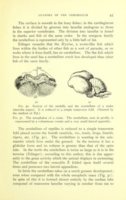
![spinal column, the fundamental fold gives rise to the motor nerves. The alar fold receives the termination of the medullary sensory nerves. The alar fold then divides into two segments: the one internal or jugal, and the other external or rhomboidal lip of His. The jugal segment becomes later on, first, in the medullary regions, the nucleus of the tract of GoU, the gray wing, the acoustic tuber- cle ; and second, in the pontine region, the locus cceruleus. The rhomboidal lip forms- in its turn first, in the medullar}^ region, the medullary or inferior olive, the accessory olivary nuclei, the nucleus of the tract of Burdach, the nuclei of the lat- eral tracts, the arcuate nuclei of the pyramids, and the gelatinous substance of Rolando. The fibers which arise from them will form the internal arcuate fibers of the medulla, the system of olivary fibers, the trapezoid body, the interolivary layer and the restiform body. Second, in the pontine region the pontine olive, the gelatinous substance of Rolando, the internal arcuate fibers, the trapezoid body and the fold of the cerebellum (]. and A. Dejerinej. The cerebellum, therefore, is developed along the path of the sensory tracts, and as an accessory of the sensory tract. At first it is a double organ, the two parts of which unite afterwards in the median line. The cerebellar fold first develops the vermis in its median portion; it is there that the first grooves to the num- ber of three or four appear towards the third month of intra- uterine life. The grooves of the cerebellar hemispheres appear during the fourth month. The cerebellum does not acquire its final form until about the fifth month. The fibers of the vermis myelinate much earlier than those in the hemispheres. A'. Couiparatke Anatomy The cerebellum follows a course in its development in the animal series parallel to that of the nervous system in general. Rudimentary in fishes it acquires its maximum of development in mammals. The cerebellum of fishes is situated behind the optic lobes and consists of an elongated appendix, adherent by its base in front, and free behind, implanted upon the sides of the spinal cord. The superior face is traversed by an antero-posterior groove.](https://iiif.wellcomecollection.org/image/b21274368_0050.jp2/full/800%2C/0/default.jpg)