Percussor stethoscope / by B. Wills Richardson.
- Richardson, Benjamin Wills.
- Date:
- [cbetween 1800 and 1899?]
Licence: Public Domain Mark
Credit: Percussor stethoscope / by B. Wills Richardson. Source: Wellcome Collection.
Provider: This material has been provided by The University of Glasgow Library. The original may be consulted at The University of Glasgow Library.
110/126 page 16
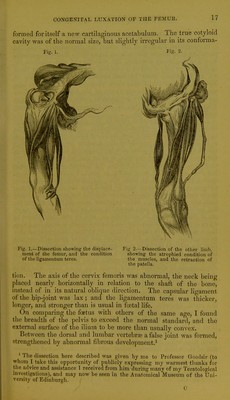
![It would seem from these statistics that the formative power (vis formativa natural) becomes weakened by being frequentlj- called into action ; and we think we may safely conclude—reasoning analogi- cally—that, in this respect, malformations by duplicity are governed by the same laws as normal twins. NO. VII. CONGENITAL LUXATION OF THE FEMUR. In the beginning of February 1853, Professor Simpson kindly gave me a malformed foetus for dissection, wliich had been sent to him by an obstetrical ]nactitioner for minute examination. On inspection I ascertained that congenital dislocation of the femur existed on both sides; and so interesting were the appearances presented ^by the dissection, that I consider the following account and delineations of them not unworthy of a place among these Contributions.' For, although this congenital displacement is by no means uncommon, opportunities of examining it in the dead body ai'e, as acknowledged by Dupuytren,^ comparatively rare, inasmuch as it is merely an infirmity, and not a disease necessarily abbreviating human life. External appearance of the Foitus.—I learned that the child had been prematurely still-born, and from its size I considered its intra- uterine life to have ended about the beginning of the eighth month of gestation. Tiie head, trunk, and upper extremities were per- fectly normal in their development; but the lower part of the body presented all the characteristic marks of congenital luxation of the femur. The limbs—considerably shortened and atrophied—were disproportioned to the size of the trunk, and obliquely placed in relation to it. They were also rotated very much inwards, and both feet were inverted by talipes vanis. The femur admitted of very little motion ; and on both sides its trochanters formed prominent projections above the site of the cotyloid cavity. The knee-joints were as immobile as if they had been anchylosed. The back could be bent abnormally between the dorsal and lumbar regions. Dissection.—On both sides the following appearances presented themselves :— The muscles of the thigh and gluteal region weie veiy much atrophied and contracted, especially where they surrounded the hip- joint. By the retraction of the atrophied and shortened rectus femoris muscle, the patella and head of the tibia were drawni up over the condyles of the femur, thereby rendering flexion of the knee- joint impossible. The head of the femur, displaced upwards and outwards, lay on the dorsum of the ilium, where it had partially * Professor Simpson did me the lionour of exhibiting my dissection to the Edinburgh Medico-Chiruigical Society, at their meeting on 16th February 1853. (See MonfJilj/Journal, vol. xvi. p. 5G7) I subsequently exhibited the preparation to tlie Edinburgh Physiological Society. (See Transactions of tlie Societi/ in Monthly Journal, vol. xvi. p. 470). ' Lea occasions de determiner, par Pouverture des coros, la nature de cette singuliere espece de luxation sont fort rares.—CTi?M<?tt<! Uhir^irgicaU, p. 82.](https://iiif.wellcomecollection.org/image/b21477784_0110.jp2/full/800%2C/0/default.jpg)