The essentials of histology : descriptive and practical for the use of students / by E.A. Schäfer.
- Edward Albert Sharpey-Schäfer
- Date:
- 1894
Licence: Public Domain Mark
Credit: The essentials of histology : descriptive and practical for the use of students / by E.A. Schäfer. Source: Wellcome Collection.
278/344 page 258
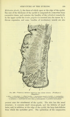
![of the nerve-fibres and their termination amongst the epithelium-cells as shown in chloride of gold preparations have been already studied (Lesson XXL). 5. Mount in Canada balsam sections of a cornea which has been stained with nitrate of silver. Notice the branched cell-spaces corresponding with the connective-tissue cells of the last preparation. [This preparation is best made by rubbing the surface of the cornea with lunar caustic after scraping off the epithelium. After ten or fifteen minutes (by which time the nitrate of silver will have penetrated the thickness of the cornea) the eye is washed with distilled water, and exposed to the light. When brown, tangential sections may be made, for which purpose the cornea may be hardened in spirit.] LESSON XLIII. L Remove the sclerotic from the anterior part of an eye which has been preserved in Miiller's fluid, and tear off thin shreds from the surface of the choroid, including amongst them portions of the ciliary muscle. Stain the shreds with htematoxylin and mount them in Farrant. Sketch the branched pigment-cells, the elastic network, the mode of attachment of the fibres of the ciliary muscle, etc. 2. Injected preparation of choroid and iris. Mount in Canada balsam portions of the choroid coat and iris from an eye, the blood-vessels of which have been filled with coloured injection. Make sketches showing the arrangement of the capillaries and veins. 3. Teased preparation of retina. Break up with needles in a drop of glycerine a minute fragment of retina which has been placed in 1 per cent, osmic acid solution for a few hours, and has subsequently been kept in dilute glycerine. Complete the separation of the retinal elements by tapping the cover-glass. Draw carefully under a high power some of the isolated elements—e.g. the rods and cones with their attached fibres and nuclei, the inner granules, the ganglion-cells, the fibres of Miiller, hexagonal pigment- cells, etc. In some of the fragments the arrangement of the elements in the retinal layers may be made out even better than in actual sections.^ Measure the length and diameter of some of the cones, the length of the cone-fibres, and the diameter of some of the outer and inner nuclei. 4. Teased preparation of lens. Separate in water the fibres of a crystalline lens which has been macerated for some days in bichromate of potash solu- tion. Sketch some of the fibres, together and separate. The eyelids (fig. 288) are covered externally by the skin, and inter- nally or posteriorly by a mucous membrane, the conjunctiva, which is reflected from them over the globe of the eye. They are composed in the main of connective tissue, which is dense and fibrous under the conjunctiva, where it forms what is known as the tarsus. Embedded in the tarsus is a row of long sebaceous glands (the 1 The distribution of the nerve-fibres and cell-processes within the retina can only be made out satisfactorily by the employment of Golgi's method (see Apipendix).](https://iiif.wellcomecollection.org/image/b20400585_0278.jp2/full/800%2C/0/default.jpg)