An atlas of skiagrams : illustrating the development of the teeth with explanatory text / by Johnson Symington and J. C. Rankin.
- Johnson Symington
- Date:
- 1908
Licence: In copyright
Credit: An atlas of skiagrams : illustrating the development of the teeth with explanatory text / by Johnson Symington and J. C. Rankin. Source: Wellcome Collection.
Provider: This material has been provided by The University of Glasgow Library. The original may be consulted at The University of Glasgow Library.
38/68 (page 22)
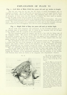
![Fig. I.—Left Side of Male Child five years old and 33', inches in height. This boy was sliuil lur his aye, l»ut his tlu>iiuL' ol denial developiueiil may he regarded as normal. All the milk teeth are in position except the u]){)er lateral incisor and the first molar, which have been lost. The remaiuins; milk teeth show no signs of absorption of their roots, but the deposit of dentine has been small in amount, leaving the pulp cavities and the canals in the roots large. The permanent teeth are all visible in the skiagram except the third molars. The crown of the lower second permanent molar is smaller than usual, and is turned almost directlv in- wards. The second bicuspids have commenced to calcify, and the first bicuspids, although more advanced in tlu'ir development, have still the crowns incompletely formed. Fig. 2.—Right Side of Boy six years old and 42 inches high. All the teeth, both temporary and permanent, in this l)eaunliil specimen are in an excellent state of preservation, and, as showm in the skiagram, they are all more or less calcified, with the exception of the third molars. The first permanent molars are erupted, and are in a line with the biting edge of the temporary teeth. The process of absorption of the roots of the milk teeth, with the possible exception of the first molars, does not a})pear to have commenced. It is somewhat unusual to find the first permanent molars so full}' erupted when the tem- porary incisors are still firmly fixed in position. The development of the roots of the first permanent molars is still incomplete, as is evident from the size of the openings at the apices of their roots. The permanent incisors and canines exhibit traces of root formation, Irat in the bicuspids and second molars calcification has not extended beyond the crown. The bony arcades over the bicuspids and separating them from the temporary molars are well seen in the mandible. The size and shape of the pulp cavities of the two lower milk molars and the first permanent molar, the canals in their roots, the thin dark lines around the roots repre- senting the thickness of the periodontal membrane, and the structure of the alveolar walls between these teeth are shown in the skiag-rams with diagrammatic clearness. The white spot .surrounded by a grey ring on the niandiMe below the roots of the first permanent molar is due to a Haw in the tihn nf tlie negative. Fig. B.—This is a view in mi tlie lateral aspect of the right maxilla, A-e., dissected in the same way as Fig. A, p. 14. The maxillary sinus is 3 cm. from before backwards, 1'7.5 cm. from above downwards, and it extends for- wards as far as a line prolonged ver- tically upwards between the first and second temporary molars, and jios- teriorly to a vertical line about 8 nun. behind the first permanent molar. Tile mieniiiL; f the sinus is situated I cm. behind the anterior end of tlie sinus and about '2 im. in front of its posterior extremity. In front the sinus reaches outwards to the infra- orbital canal, but behind it extends about 4 mm. external to it, and lies above the first and second permanent molars, its floor being about '2 cm. above the grinding surface of the first permanent molar.](https://iiif.wellcomecollection.org/image/b21458868_0038.jp2/full/800%2C/0/default.jpg)