An atlas of skiagrams : illustrating the development of the teeth with explanatory text / by Johnson Symington and J. C. Rankin.
- Johnson Symington
- Date:
- 1908
Licence: In copyright
Credit: An atlas of skiagrams : illustrating the development of the teeth with explanatory text / by Johnson Symington and J. C. Rankin. Source: Wellcome Collection.
Provider: This material has been provided by The University of Glasgow Library. The original may be consulted at The University of Glasgow Library.
42/68 (page 26)
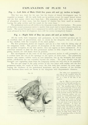
![Fig. I. Right Side of Boy seven years old and 44 inches high. The erupted teeth of tlie maxilla are the ]MTin,iiiciit ceniral incisor, teiiiporarv canine, first and second temporarv and lir-i ]n'liiiaiirnt molar: l)otli nulk incisors have therefore been shed, luit oidy one permanent incisor — the central—is throuuh the uum. The three temporary teeth still persisting are moio or less carious. The skiagram shows five permanent teeth cmhedded in the maxilla. \\/.. the lateral incisor, the canine, the first and second bicuspid, and the second molar. The i-oots of the temporary molars are almost entirely absorbed. This specimen shows clearlv the hish position of the permanent canine as compared with the neighbouring non-erupted per- manent teeth, and the irregularity of the dental arch which would arise should the canine begin to descend before the lateral incisor and the Hi'st bicusphl ha\c crupied, ami li\- ihcii' divergence during the process left room for the canine. There is no trace of a third molar. The teeth of the mandible correspond to those of the maxilla, except that the lateral milk incisor is still in position. Both the temporary molars are afiected with caries. Fig. C is a drawing of tlie lateral a-spect of this .si)ecimc'n di.-<sected to sbow tlic liniiiir iiRMiilirane of the maxillaiy aiitrHin and othvr accessory nasal .sinuses. This antrum is slij.rbtly larger in all directions than that of tlic boy six years old, being 3-4 cm. frimi before backwards. 2 cm. from al)o\e downwards, and TS cm. from within outwards. Its floor presents posteriorly a prominence opposite the socket for the second permanent molar, and tlic bone is so thin here that it can lie easily pierced with a needle. A little in fnuit iif this prominence there is another situated above the alveolus of the second bicuspid. The depression between these two prominences is above the first iiermanent molar. Opposite the first bicuspid tlu' sinus turns sharply upwards on to tlie anterior wall, wliicli has a distinct ]irMiniiicnce due to the pernument raniiic timtli. Fig. 2.—Left Side of same Boy, The upper dental arch difi'ers from that of the right side, as the tein]M)iai\- lateral incisor is still in position, while the first temporary molar is shed and the first bicuspid erupted. 'J'he ap])earance through the gum of the first bicuspid is obviously premature, as its root is only just beginning to foi'm. The second temporary molar has lost the greater part of its roots, and the second bicuspid, situated just above it, has attaincil to nearly the same stage of development as the first bicuspid. The condition of the teeth in this half of the mandible closely corresponds to that of the right side, all the milk teeth being in position except the central incisor. 2(5](https://iiif.wellcomecollection.org/image/b21458868_0042.jp2/full/800%2C/0/default.jpg)