Licence: Public Domain Mark
Credit: Elements of histology / by E. Klein. Source: Wellcome Collection.
Provider: This material has been provided by the Royal College of Physicians of Edinburgh. The original may be consulted at the Royal College of Physicians of Edinburgh.
115/376 (page 99)
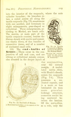
![meshes. Thus the pei-ipheral third or fourth of the gland is subdivided by tlie septa and trabeculse, into relatively large spherical or oblong compartments, while the middle portion is made up of relatively small cylin- drical or irregularly-shaped compartments (I'ig. 60). The former region is the cortex, the latter the medulla of the gland. The compartments of the cortex anastomose with one another and with those of the medulla, and these latter also form one intercommunicating system. The fibrous capsule, the septa and trabeculse are the carriers of the vascular trunks ; the trabeculag consist of fibrous connective tissue and of a certain Fig. 60.—From a Vertical Section through a Lymphatic Gland, the Lymphatics of which had been injected. c, The outer capsule, with Ijmpliatic vessels in section: a, the cortical hnnph i„] I'', cortical lymph slnuse..; h, the raeduilary: injected lymph sinuses between the masses of adenoid tissue. (Atlas.) amount of non-striped muscular tissue, which is con- spicuous in some animals—e.^., pig, calf, rabbit, guinea-pig—but is scarce in man. Sometimes coarsely granular connective tissue cells](https://iiif.wellcomecollection.org/image/b21725330_0115.jp2/full/800%2C/0/default.jpg)