Licence: Public Domain Mark
Credit: Elements of histology / by E. Klein. Source: Wellcome Collection.
Provider: This material has been provided by the Royal College of Physicians of Edinburgh. The original may be consulted at the Royal College of Physicians of Edinburgh.
65/376 (page 49)
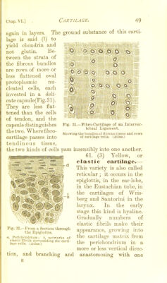
![The ground substance of this carti- to II r4 1 nu- each deli- 0' o o c o I-'. Q Fig. 31. again in layers, lage is said C?) yield chondrin and not glutin. Be- tween the strata of the fibrous bundles are rows of more or less flattened oval ])rotoplasmic cleated cells, invested in a cate capsule(Fig. 31). They are less flat- tened than the cells of tendon, and the capsule distinguishes thetwo. Wherefibro- cartilage passes into tendinous tissue, the two kinds of cells pass insensibly into one another. 61. (3) Yellow, or elastic cartilage.— This variety is also called reticular ; it occurs in the epiglottis, in the ear-lobe, in the Eustachian tube, in the cartilages of Wris- berg and Santorini in the larynx. In the early stage this kind is hyaline. Gradually numbers of elastic fibrils make their appearance, growing into the cai-tilage matrix from the perichondrium in a more or less vertical direc- -Fibro-Cartilage of an Interver- tebral Ligament. Showing the bundles of fibrous tissue and rows of cartilage cells. (Atlas.) Fig. 32.—From a Section tiirougli the Epiglottis. a, Perichondriiirn; h, networks of elastic nbrils surrounding the carti- lage cells. (Atlas.) tion, and branching and anastomosing with one](https://iiif.wellcomecollection.org/image/b21725330_0065.jp2/full/800%2C/0/default.jpg)