The incidence and bacteriological characteristics of tuberculous infection in children / by Arthur Eastwood, M.D., and Fred Griffith, M.B. An enquiry, based on a series of autopsies, into the occurrence and distribution of turberculous infection in children, and its relation to the bovine and the human types of tubercle bacilli respectively / by A. Stanley Griffith, M.D.
- Date:
- 1914
Licence: Public Domain Mark
Credit: The incidence and bacteriological characteristics of tuberculous infection in children / by Arthur Eastwood, M.D., and Fred Griffith, M.B. An enquiry, based on a series of autopsies, into the occurrence and distribution of turberculous infection in children, and its relation to the bovine and the human types of tubercle bacilli respectively / by A. Stanley Griffith, M.D. Source: Wellcome Collection.
21/188 page 13
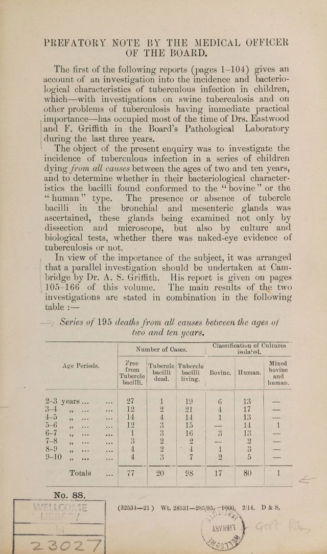
![bacilli could be found in two smears. General tuberculosis was produced in three guinea-pigs. Lungs.—Report received:—‘‘ The pleura was everywhere covered with tubercles and there were small patches, some show- ing early. caseation, throughout both lungs. Very tough pleural adhesions were present at the apices of both lungs and both apices were the seat of large caseating masses.” Mesenteric Glands.— Formed a mass, 9 x 7 x 2°59 cm., con- sisting of large adherent glands composed practically throughout of firm, opaque, yellow ish white, caseous tissue. No tubercle bacilli were found in three smears. General tuberculosis was produced in three guinea-pigs. Other Organs.—Report received :—‘‘ The peritoneum was everywhere studded with smal] tubercles. Tuberculous ulcers were present in the small intestines. There were a few scattered tubercles at the base of the brain and along the middle cerebral arteries.” Summary. Direct cultures from the cervical and bronchial glands and cultures from the mesenteric gland through a guinea-pig were all eugonic. Tested on rabbits, the bronchial and mesenteric gland cultures were of low virulence. Evidence as to portal of entry inconclusive. HAW. G. S., 6 years; male.—Morbus cordis. Iungs.—There was a brownish caseous nodule, about the size of a millet seed, in which no tubercle bacilli were seen. Spleen.—On section there were two fibrous nodules, less than rape seed in size, in which no tubercle bacilli were seen. Three guinea-pigs were found healthy six weeks after inoculation. Iaver.—On the surface was a single grey, submiliary tubercle with an opaque centre. Two guinea-pigs were found healthy SIX weeks after inoculation. | Other organs.—No visible tuberculosis. Three guinea-pigs inoculated with bronchial glands and three ico with mesenteric glands were all found healthy six weeks afterwards. Summary. Cultures from two guinea-pigs inoculated with mesenteric glands, from one inoculated with spleen, and from one inoculated with liver tubercle all remained sterile. Origin of infection probably respiratory. thao. N. D., 64 years; female. Tuberculous meningitis; clinically, no definite physical signs in chest or abdomen. Bronchial Glands.—At the bifurcation of the trachea these were caséating and ulcerating into the left bronchus; there was also a caseous gland just above the bifurcation. A few lone beaded bacilli were seen. The glands at the roots of the lungs were not enlarged and not visibly tuberculous. General tuberculosis was produced in three guinea-pigs; two others, inoculated with anti- forminised tissue, and a rabbit remained healthy.](https://iiif.wellcomecollection.org/image/b29012971_0021.jp2/full/800%2C/0/default.jpg)