The incidence and bacteriological characteristics of tuberculous infection in children / by Arthur Eastwood, M.D., and Fred Griffith, M.B. An enquiry, based on a series of autopsies, into the occurrence and distribution of turberculous infection in children, and its relation to the bovine and the human types of tubercle bacilli respectively / by A. Stanley Griffith, M.D.
- Date:
- 1914
Licence: Public Domain Mark
Credit: The incidence and bacteriological characteristics of tuberculous infection in children / by Arthur Eastwood, M.D., and Fred Griffith, M.B. An enquiry, based on a series of autopsies, into the occurrence and distribution of turberculous infection in children, and its relation to the bovine and the human types of tubercle bacilli respectively / by A. Stanley Griffith, M.D. Source: Wellcome Collection.
38/188 page 30
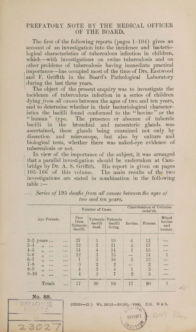
![Sr. E. G., 4 years; female. Measles and whooping cough followed by pulmonary tuberculosis and empyema. Cervical Glands.—Enlarged and beset with small caseous areas. Bronchial Glands.—Enlarged and showed chronic fibro-caseous areas; tubercle bacilli were moderately numerous. General tuberculosis was produced in three guinea-pigs. Lungs.—The left lung was solid and riddled with caseo- necrotic nodules, many softening and becoming purulent; cavities were seen in early and later stages of formation. The right lung was less severely affected; caseo-necrotic nodules and tubercles were distributed throughout. Caseo-pus from the left lung was swarming with masses of tubercle bacilli. Mesenteric Glands.—Slight enlargement; many showed dis- crete, grey and caseous tubercles. Tubercle bacilli were scanty. General tuberculosis was produced in three guinea-pigs. Spleen.—Numerous minute grey tubercles seen under capsules and on ‘section. Kidneys.—A few tubercles in cortices and medulla. Intestines.—Peyer’s patches congested ; tuberculous ulcers seen in two. Summary. Cultures, through guinea-pigs, of mesenteric and bronchial glands were eugonic and of low virulence for rabbits. Origin of infection probably respiratory. H 83. C. H., 24 years; female. Tuberculous knee-joints; operation ; tuberculous meningitis. Bronchial Glands.—At the root of the right lung and within the bifurcation were three moderately enlarged glands consisting almost entirely of caseo-caleareous material. The left glands were not affected. In the caseous material a few rather degene- rate tubercle bacilli were seen. General tuberculosis was pro- duced in one guinea-pig, the other two guinea-pigs inoculated being found free from tuberculosis. Lungs.—At the junction of the right upper and middle lobes was a pea-sized nodule with a firm fibrous capsule and a caseo- calcareous centre. Scattered throughout both lungs were moderately numerous minute, translucent tubercles. Two. euinea-pigs inoculated with the nodule, which showed moderately numerous tubercle bacilli, were icc free from tuberculosis when killed six weeks afterwards, and a culture made from one remained sterile. ‘ Mesenteric Glands.—Slightly enlarged; no visible tuberculosis, but tuberculosis was produced in two out of three guinea-pigs inoculated. Other Organs.—No visible tubercles in spleen, kidneys, liver, and intestines. Summary. Cultures from the bronchial] and mesenteric glands, through guinea-pigs, were eugonic and of low virulence for rabbits. Origin of infection probably respiratory.](https://iiif.wellcomecollection.org/image/b29012971_0038.jp2/full/800%2C/0/default.jpg)