Textbook of anatomy. Section 2. The muscular system: the nervous system: the organs of sense and integument / By D.J.Cunningham,Robert Howden and A.M.Patterson.
- Cunningham, D. J.
- Date:
- 1906
Licence: In copyright
Credit: Textbook of anatomy. Section 2. The muscular system: the nervous system: the organs of sense and integument / By D.J.Cunningham,Robert Howden and A.M.Patterson. Source: Wellcome Collection.
Provider: This material has been provided by UCL Library Services. The original may be consulted at UCL (University College London)
25/522 (page 319)
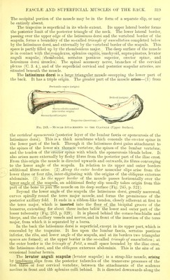
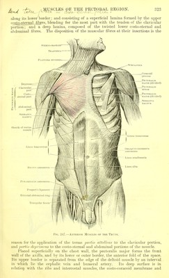
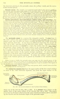
![The occipital portion of the muscle may be in the form of a separate slip, or may be entirely absent. The trapezius is superficial in its whole extent. Its upper lateral border forms the posterior limit of the posterior triangle of the neck. The lower lateral border, passing over the upper edge of the latissimus dorsi and the vertebral border of the scapula, forms a boundary of the so-called triangle of auscultation completed below by the latissimus dorsi, and externally by the vertebral border of the scapula. This space is partly filled up by the rhomboideus major. The deep surface of the muscle is in contact with the complexus, splenius capitis, omohyoid, supraspinatus, levator anguli scapulae, rhomlDoidei, serratus posticus superior, erector spina?, and latissimus dorsi muscles. The spinal accessory nerve, branches of the cervical plexus (C. 3. 4.), and of the superficial cervical and posterior scapular arteries, are situated beneath the muscle. The latissimus dorsi is a largejriangular muscle occupying the lower part of the back. It has a triple origin. The greater part of the muscle arises—(1) from Pectoralis major (origin) Fig. 243.—Muscle-Attachments to the Clavicle (Upper Surface). the vertebral aponeurosis (posterior layer of the lumbar fascia or aponeurosis of the latissimus dorsi). This is a thick membrane which conceals the erector spin« in the lower part of the back. Through it the latissimus dorsi gains attachment to the spines of the lower six thoracic vertebrae, the spines of the lumbar vertebrae, and the tendon of the erector spinae with which the aponeurosis blends below. It also arises more externally by fleshy fibres from the posterior part of the iliac crest. From this origin the muscle is directed upwards and outwards, its fibres converging to the lower angle of the scapula. In relation to its upper and outer borders additional fibres arise. (2) Along the outer border muscular slips arise from the lower three or four ribs, inter-digitating with the origins of the obliquus externus abdominis. (3) As the upper border of the muscle passes horizontally over the lower angle,of the scapula, an additional fleshy slip usually takes origin from this part of the boneTojoin tlie muscle on its deep surface (Fig. 245, p. 321). Beyond the lower angle of the scapula the latissimus dorsi, greatly narrowed, curves spirally round the teres major muscle, and forms the prominence of the posterior axillary fold. It ends in a ribbon-like tendon, closely adherent at first to the teres major, which is inserted into the floor of the bicipital groove of the humerus, extending for about three inches below the lower and outer part of the lesser tuberosity (Fig. 253, p. 329). It is placed behind the coraco-brachialis and biceps, and the axillary vessels and nerves, and in front of the insertion of the teres major, from which it is separated by a bursa. In the back the latissimus dorsi is superficial, except in its upper part, which is concealed by the trapezius. It lies upon the lumbar fascia, serratus posticus inferior, the ribs, and lower angle of the scapula, and at its borders two triangular spaces are formed; at the upper border is the so-called triangle of auscultation; at the outer border is the triangle of Petit, a small space bounded by the iliac crest, the latissimus dorsi, and the obliquus externus abdominis. This is the site of an occasional lumbar hernia. The levator anguli scapulae (levator scapulae) is a strap-like muscle, arising by tendinous sli]>s horn, the posterior tubercles of the transverse processes of the first three or lour cervical vertebrae, between the attachments of the scalenus niedius in front and tli*e splenius colli behind. It is directed downwards along the](https://iiif.wellcomecollection.org/image/b21271070_0025.jp2/full/800%2C/0/default.jpg)