Demonstrations of anatomy : being a guide to the knowledge of the human body by dissection / by George Viner Ellis.
- George Viner Ellis
- Date:
- 1856
Licence: Public Domain Mark
Credit: Demonstrations of anatomy : being a guide to the knowledge of the human body by dissection / by George Viner Ellis. Source: Wellcome Collection.
Provider: This material has been provided by King’s College London. The original may be consulted at King’s College London.
54/856 page 38
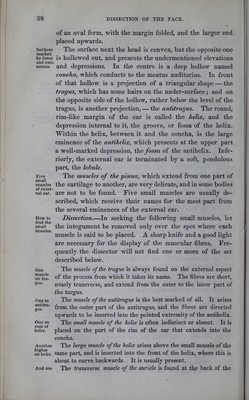
![Surfaces marked by fossJE and emi- nences. Five small muscles of exter- nal ear. How to find the small muscles. One muscle on tra- gus. One to antitra- gus. One on root of helix. Another higher on helix. And one of an oval form, with the margin folded, and the larger end placed upwards. The surface next the head is convex, but the opposite one is hollowed out, and presents the undermentioned elevations and depressions. In the centre is a deep hollow named concha, which conducts to the meatus auditorius. In front of that hollow is a projection of a triangular shape — the tragus, which has some hairs on the under-surface; and on the opposite side of the hollow, rather below the level of the tragus, is another projection, — the antitragus. The round, rim-like margin of the ear is called the helix, and the depression internal to it, the groove, or fossa of the helix. Within the helix, between it and the concha, is the large eminence of the antihelix, which presents at the upper part a well-marked depression, the fossa of the antihelix. Infe- rior] y, the external ear is terminated by a soft, pendulous part, the lobule. The muscles of the pinna, which extend from one part of the cartilage to another, are very delicate, and in some bodies are not to be found. Five small muscles are usually de- scribed, which receive their names for the most part from the several eminences of the external ear. Dissection.—In seeking the following small muscles, let the integument be removed only over the spot where each muscle is said to be placed. A sharp knife and a good light are necessary for the display of the muscular fibres. Fre- quently the dissector will not find one or more of the set described below. The muscle of the tragus is always found on the external aspect of the process from which it takes its name. The fibres are short, nearly transverse, and extend from the outer to the inner part of the targus. The muscle of the antitragus is the best marked of all. It arises from the outer part of the antitragus, and the fibres are directed upwards to be inserted into the pointed extremity of the antihelix. The small muscle of the helix is often indistinct or absent. It is placed on the part of the rim of the ear that extends into the concha. The large muscle of the helix arises above the small musele of the same part, and is inserted into the front of the helix, where this is about to curve backwards. It is usually present. The transverse muscle of the auricle is found at the back of the](https://iiif.wellcomecollection.org/image/b21308597_0054.jp2/full/800%2C/0/default.jpg)