An American text-book of diseases of the eye, ear, nose and throat / edited by G.E. DeSchweinitz and B. Alex. Randall.
- Date:
- 190l
Licence: Public Domain Mark
Credit: An American text-book of diseases of the eye, ear, nose and throat / edited by G.E. DeSchweinitz and B. Alex. Randall. Source: Wellcome Collection.
57/700 page 53
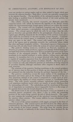
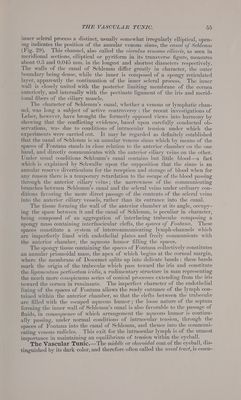
![anterior layers first having joined the conjunctival nerves before proceeding to the cornea. The more numerous branches which pass directly to the corneal stroma from the annular plexus enter the substantia propria near the _ posterior limiting membrane, the far greater number, however, passing to the anterior lamella, only about one-third of the nerves which enter the cornea being distributed to the posterior layers. The nerve-bundles, on penetrating into the corneal stroma, are invested for a short distance, from 0.75—] mm., by perineural lymph-sheaths, the individual nerve-fibers losing their medul- lary sheaths at about the same time. After entering the substantia propria the nerves form the fwndamental plexus within the corneal stroma, from which numerous lateral branches are given off at various levels; these are composed of non-medullated fibers which soon break up into the component varicose fibrille. In addition to the lateral twigs, perforating branches ascend through the anterior lamellz as far as the epithelium, beneath which they form the subepithelial plexus. The terminal fibers of this plexus in many instances enter the epithelium to end either in special end-bulbs or between the cells as the intra-epithelial plexus. The plexuses within the substantia propria formed by the twigs given off at various levels spread out between the lamelle of fibrous tissue; the nodal points or places of meeting of the fibers are often marked by angular areas outlined by the interlacing fibers; nuclei, belonging to the delicate nerve- sheaths, are sometimes present. The terminal fibers of the corneal nerves are related to various forms of end-organs, among which are intricate convo- lutions, less-contorted loops and hooks, and irregular quadrate plates. The Sclera.—The sclerotic coat forms the posterior four-fifths of the fibrous tunic of the eyeball, contributing largely to the protection and sup- port of the more delicate structures within, as well as affording the points of attachment of the ocular muscles. Although composed of practically the same histological elements as the cornea, the disposition of these is such that the dead-white opacity is produced which so conspicuously contrasts with the beautifully transparent cornea. 3 The sclera is thickest over the posterior third of the ball, where the maintenance of a uniform curvature for the support of the retina is of great importance: in the vicinity of the optic nerve the sclerotic coat measures nearly 1 mm. in thickness, gradually becoming thinner toward the anterior boundary, until beneath, or just posterior to, the zone of attachment of the recti muscles the sclera is reduced to about 0.4 mm. Anterior to the tendon- zone the thickness of the fibrous tunic is augmented by the expansion of the muscle insertions until it reaches about 0.6 mm. In individuals possessing thin sclerze and deeply pigmented eves the sclerotic coat presents a bluish or skimmed-milk tint, due to the deeply-colored tissue beneath the fibrous coat ; this bluish appearance is well marked in the eyes of young children. In its structure the sclera closely resembles the cornea, being composed of interlacing bundles of fibrous tissue disposed with much greater irregu- larity, however, than those of the cornea. The clefts between the fibrous bundles correspond to the corneal spaces and contain irregularly stellate connective-tissue cells—the scleral corpuscles. The scleral spaces are less regularly arranged and possess a less elaborate system of connecting canal- iculi. The scleral bundles further differ from those of the cornea in contain- ing numerous elastic fibers and in yielding gelatin on boiling: their general disposition is equatorial and meridional, although the bundles interlace with one another at all angles. In addition to the usual branched scleral corpuscles, those occupying the](https://iiif.wellcomecollection.org/image/b29003064_0057.jp2/full/800%2C/0/default.jpg)
 No text description is available for this image
 No text description is available for this image
 No text description is available for this image