Volume 1
Diseases of the organs of respiration : a treatise on the etiology, pathology, symptoms, diagnosis, prognosis, and treatment of diseases of the lungs and air-passages / by Samuel West.
- West, Samuel (Samuel Hatch), 1848-1920.
- Date:
- 1902
Licence: Attribution-NonCommercial 4.0 International (CC BY-NC 4.0)
Credit: Diseases of the organs of respiration : a treatise on the etiology, pathology, symptoms, diagnosis, prognosis, and treatment of diseases of the lungs and air-passages / by Samuel West. Source: Wellcome Collection.
19/420 (page 3)
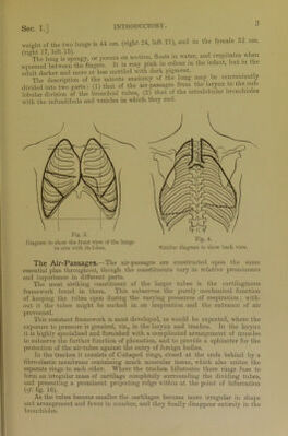
![Sec ]..] weight of the two lungs is 44 ozs. (right 24, left 21), and in the female 32 ozs. (rigThe lui^is spongy, or porous on section, floats in water, and crepitates when squeezed betwemfthffingers. It is rosy pink in colour in the infant, but in the adult darker and more or less mottled with dark pigment. The description of the minute anatomy of the lung may be conveniently divided into two parts : (1) that of the air-passages from the larynx to the sub- lobular division of the bronchial tubes, (2) that of the intralobular bronchioles with the infundibula and vesicles in which they end. Fig. 3. Diagram to show the front view of the lungs in silu with its lobes. Fig. 4. Similar diagram to show back view. The Air-Passages.—The air-passages are constructed upon the same essential plan throughout, though the constituents vary in relative prominence and importance in different parts. The most striking constituent of the larger tubes is the cartilaginous framework found in them. This subserves the purely mechanical function of keeping the tubes open during the varying pressures of respiration; with- out it the tubes might be sucked in on inspiration and the entrance of air prevented. This resistant framework is most developed, as would be expected, where the exposure to pressure is greatest, viz., in the larynx and trachea. In the larynx it is highly specialised and furnished with a complicated arrangement of muscles to subserve the further function of phonation, and to provide a sphincter for the protection of the air-tubes against the entry of foreign bodies. In the trachea it consists of C-shaped rings, closed at the ends behind by a fibro-elastic membrane containing much muscular tissue, which also unites the separate rings to each other. Where the trachea bifurcates these rings fuse to form an irregular mass of cartilage completely surrounding the dividing tubes, and presenting a prominent projecting ridge within at the point of bifurcation (cf. fig. 16). As the tubes become smaller the cartilages become more irregular in shape and arrangement and fewer in number, and they finally disappear entirely in the bronchioles.](https://iiif.wellcomecollection.org/image/b28121909_0001_0019.jp2/full/800%2C/0/default.jpg)