Volume 1
A system of anatomical plates of the human body; accompanied with descriptions, and physiological, pathological & surgical observations. Text and plates / By John Lizars.
- John Lizars
- Date:
- [1840?]
Licence: Public Domain Mark
Credit: A system of anatomical plates of the human body; accompanied with descriptions, and physiological, pathological & surgical observations. Text and plates / By John Lizars. Source: Wellcome Collection.
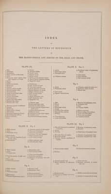
496/535 page 5
![| ) l, Smooth surface of linea aspera, made by superficial femoral artery and vein m, Internal condyle p Tubercle giving origin to fibular d e head of gastrocnemius exter- nus muscle r, Surface giving origin to tibial head of gastrocnemius exter- nus muscle t, Cavity or notch, to which are attached crucial ligaments a, Superior fibular division of linea to gluteus maximus muscle ¢, Point of attachment of gluteus medius muscle a. Point of insertion of rectus and erureus muscles b, Point of insertion of vastus in- ternus muscle c, Point of insertion of vastus ex- ternus muscle d, Point of attachment of patellar ligament d, Point of attachment of patellar ligament e, Point of attachment of capsular ligament m, Surface opposed to internal con- dyle of os femoris n, Surface opposed to external con- dyle of os femoris a, Body or shaft of tibia d, Tubercle of tibia e, Surface into which inner ham- string muscles are inserted h, Surface to which fibula is arti- culated g, Styloid process or malleolus in- ternus of tibia m, Fibular angle affording attach- ment to interosseous lga- ment _y, Smooth articular surface for as- tragalus tachment to fibula 1, Exterior angle of body or shaft of fibula 2, Proximal extremity of fibula 4, Tibial angle giving attachment to interosseous ligament 9, Distal extremity of fibula 10, Coronoid process or malleolus externus 11, Smooth surface opposed to as- tragalus 6, Hievation on head of tibia, giving attachment tocrucial ligaments jf, Point of insertion of semi-mem- branosus muscle tibia crucial ligament k, Oblique ridge made by gastroc- nemius internus muscle m, Fibular angle giving insertion to interosseous ligament n, Groove made by flexor longus digitorum pedis and _ tibialis posticus muscles 0, Posterior tibial angle made by tibialis posticus muscle, &c. p, Groove made by flexor longus muscle gq, Styloid process or malleolus in- ternus a, Surface opposed to fibula a, Os astragalus 6, Os naviculare e, Os calcis d, Os cuneiforme internum é, Os cuneiforme medium Ff, Os cuneiforme externum g, Os cuboides k, Depression made by flexor longus pollicis pedis m, Smooth projection of astragalus nm, Smooth surface of naviculare, where it joins cuboides t, Projection of metatarsal bone of little toe, affording insertion to peroneus brevis muscle ¢, Smooth surface of os calcis op- posed to os cuboides, g gq, Fossa made by peroneus longus muscle 1, Situated on os naviculare, 6, in- dicates surface opposed to os cuneiforme internum, d@ 1, Metatarsal bone of great toe 2, Situated on os naviculare, J, in- dicates surface opposed to os cuneiforme medium, ¢ 2, Situated on cs cuneiforme ex- ternum, f, indicates surface v 1, Exterior angle of body or shaft of fibula 2, Proximal extremity of fibula 3, Foramen for nutritious vessels 4, Tibial angle giving attachment to interosseous ligament 6, Surface giving origin to fibular head of gastrocnemius inter- nus muscle 9, Distal extremity of fibula 10, Coronoid process or malleolus externus 12, Cavity for mucilaginous glands 13, Groove made by peronei mus- cles opposed to metatarsal bone, 2, of index toe 2, Metatarsal bone of index toe 3, Situated on os naviculare, J, in- dicates surface opposed to os cuneiforme externum, f 3, Situated on os cuneiforme ex- ternum, jf, indicates surface opposed to metatarsal bone, 3, of middle toe 3, Metatarsal bone of middle toe 4, Situated on os cuneiforme ex- ternum, f, indicates surface opposed to metatarsal bone of ring toe, 4 4, Metatarsal bone of ring toe 5, Metatarsal bone of little toe 6, First bone of great toe 7, Last bone of great toe 8, First bone of index toe 9, Second bone of index toe 10, Third bone of index toe ]1, First bone of middle toe 12, Second bone of middle toe 13, Third bone of middle toe 14, First bone of ring toe 15, Second bone of ring toe 16, Third bone of ring toe 17, First bone of little toe 18, Second bone of little toe 19, Third bone of little toe ® 1} | | |](https://iiif.wellcomecollection.org/image/b33543008_0001_0496.jp2/full/800%2C/0/default.jpg)

 No text description is available for this image
 No text description is available for this image
 No text description is available for this image