The essentials of histology : descriptive and practical for the use of students / by E.A. Schäfer.
- Edward Albert Sharpey-Schäfer
- Date:
- 1898
Licence: Public Domain Mark
Credit: The essentials of histology : descriptive and practical for the use of students / by E.A. Schäfer. Source: Wellcome Collection.
291/376 page 279
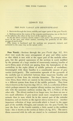
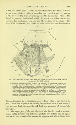
![LESSON XLI. THE PONS VAROLII AND MESENCEPHALON 1. Sections through the lower, middle, and upper parts of the pons Varolii. 2. Sections across the region of the corpora quadrigemina, one at the level of the inferior, the other at the level of the superior, pair. In all the above sections sketch under a low power the general arrange- ment of the grey and white matter, inserting the positions of the chief groups of nerve-cells. [The tissue is hardened and the sections are prepared, stained, and mounted in the same way as the spinal cord.] Pons Varolii.—Sections through the pons Varolii (figs. 317, 318) show very much the same arrangement of grey and white matter as that which is met with at the upper part of the medulla oblon- gata, but the general appearance of the sections is much modified by the presence of a large number of transversely coursing bundles of nerve-fibres, most of which are passing from the hemispheres of the cerebellum to the raphe (fibres of middle peduncle of cerebellum). Intermingled with these bundles is a considerable amount of grey matter (nuclei pontis). The continuation upwards of the pyramids of the medulla (py) is embedded between these transverse bundles and separated by them from the reticular formation. The deeper trans- verse fibres, those which are nearest to the reticular formation, belong- to a different system from those of the middle peduncle. They form what is known as the trapezium (figs. 317, tr.; 318, ^); a collection of fibres which perhaps connects the superior olivary nucleus (see below) of one side with the accessory auditory nucleus (fig. 316, n.VIIIac.) of the other side. The olivary nucleus is no longer seen, but there are one or two small collections of grey matter much more conspicuous in some animals than in man, which lie in the ventral part of the reticular formation, and are known as the superior olivary nucleus (o.s). Another important collection of large nerve-cells which is found in the upper part of the medulla oblongata, and extends into the pons Varolii, lies near the floor of the fourth ventricle, a little mesial to the restiform body : this is known as the nucleus of Deiters (D, fig. 317). The nerve- fibres connected with its cells are stated to be continued downwards](https://iiif.wellcomecollection.org/image/b20400986_0295.jp2/full/800%2C/0/default.jpg)