Surgical pathology and principles / by J. Jackson Clarke.
- Clarke, J. Jackson (James Jackson), 1860-1940.
- Date:
- 1897
Licence: Public Domain Mark
Credit: Surgical pathology and principles / by J. Jackson Clarke. Source: Wellcome Collection.
64/482 (page 42)
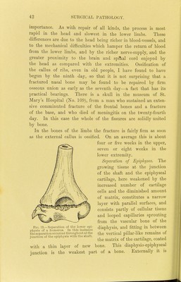
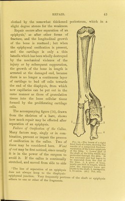
![importance. As with repair of all kinds, the process is most rapid in the head and slowest in the lower limbs. These differences are due to the head being richer in blood-vessels, and to the mechanical difficulties which hamper the return of blood from the lower limbs, and by the richer nerve-supply, and the greater proximity to the brain and sjjitial cord enjoyed by the head as compared with the extremities. Ossification of the callus of ribs, even in old people, I have found to have begun by the ninth day, so that it is not surprising that a. fractured nasal bone may be found to be repaired by firm osseous union as early as the seventh day—a fact that has its practical bearings. There is a skull in the museum of St. Mary's Hospital (No. 109), from a man who sustained an exten- sive comminuted fracture of the frontal bones and a fracture of the base, and who died of meningitis on the twenty-fourth day. In this case the whole of the fissures are solidly united by bone. In the bones of the limbs the fracture is fairly firm as soon as the external callus is ossified. On an average this is about four or five weeks in the upper, seven or eight weeks in the lower extremity. Separation of Einpliyses. The growing tissue at the junction of the shaft and the epiphyseal cartilage, here Aveakened by the increased number of cartilage cells and the diminished amount of matrix, constitutes a narrow layer with parallel surfaces, and consists partly of cellular tissue and looped capillaries sprouting from the vascular bone of the diaphysis, and fitting in between the separation Ofcurreii tliroug]iout at the vertical piUar-llke rcmamS 01 junction of tliecpipliysis with the shaft. . i,, i. j the matrix of the cartilage, coated with a thin layer of new bone. This diaphysio-epiphyseal junction is the weakest part of a bone. Externally it is Fid. 13.—Separation of tlie lower epi pliysis of a humerus. In tliis instance](https://iiif.wellcomecollection.org/image/b20412290_0064.jp2/full/800%2C/0/default.jpg)