Principles of surgery / By N. Senn ... Illustrated with 109 wood-engravings.
- Nicholas Senn
- Date:
- 1890
Licence: Public Domain Mark
Credit: Principles of surgery / By N. Senn ... Illustrated with 109 wood-engravings. Source: Wellcome Collection.
Provider: This material has been provided by the Augustus C. Long Health Sciences Library at Columbia University and Columbia University Libraries/Information Services, through the Medical Heritage Library. The original may be consulted at the the Augustus C. Long Health Sciences Library at Columbia University and Columbia University.
32/670 page 12
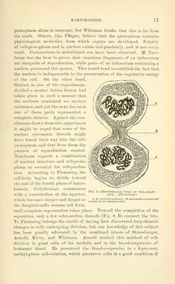
![the solution is kept at a iJiopcr tcniperature in the moist chaml)er on the o])ject-u,hiss. If to tills solution a 25-per-cent. solution of chloride of U'old is added, the karyokinetic figures are made clearer. In studying the process of karyokinesis in fixed tissue-cells in a state of infiltration, it is necessary to resort to the fixation and staining methods described by Flemming. The modern observers who have studied regeneration of epithelial cells have come to the conclusion that cell-division takes place almost exclusively by karyokinesis. Podwyssozki has studied this method of cell-reproduction with special reference to regeneration of liver-cells, and has come to some ver}' important conclusions. In cats and young guinea-pigs he observed, after injurj' of the liver, extra-nuclear chromatin substance before he could detect any karyokinetic figures in the nucleus. The chromatin in the cell-body appeared in two forms,-— either as fine granules scattered diffusely through the protoplasm of the cell, or a,s lumps of chromatin, and he designated these larger masses as procliromatin ; but he also noticed that the granular form, at a later stage, aggregated and formed masses which united with the nuclear chromatin. Klebs explains the presence of chromatin in the cell-protoplasm to an extra-cellular origin,—the leucocytes. He believes that the chromatin contained in leucocytes is liberated after fragmentation has taken place and enters the 3'oung cells, where they serve as food and become a part of the nuclear net-work. This view is strengthened by the statement of Podwyssozki tiiat he found numerous leucoC3'tes in the immediate vicinity of the new cells. Ziegler and Obolensk}'produced arsenical intoxication in animals l)y administering in daily doses subcutaneously, and when they examined the liver they found well-marked karyokinetic figures in the endothelial cells of the intra-acinous capillaries, the epithelia of the bile-ducts, and, less frequently, in the secreting cells. Karyokinetic figures were first visible in the nuclei of the capilhuy endothelia, and Avere undoubtedl3' caused by the direct action of the arsenic upon the cells. These experiments show that karyokinesis will follow the applica- tion of chemical as well as traumatic irritants. FRAGMENTATION OF NUCLEUS Arnold and Pfitzner have described, in giant and other cells under- going pathological changes, direct fragmentary division of the nucleus, by which it may break up into many parts, often of unequal size, without contemporaneous division of the cell. Arnold and others have also de- scribed incomplete fragmentation of the nucleus where the nuclear masses remain connected with each other, and can be seen as lobulated and reticulated structures. Arnold saw fragmentation of the nucleus in the cells of the marrow of bone and in leucocytes undergoing transformation](https://iiif.wellcomecollection.org/image/b21207501_0032.jp2/full/800%2C/0/default.jpg)