Papilliform lesions (lymphangiomata) of the scrotum : associated with multiple petechial spots on the trunk and limbs : being a paper read in the Section of Dermatology at the Annual Meeting of the British Medical Association, Liverpool, 1912 / by Frank Cole Madden.
- Madden, Frank Cole, 1873-1929.
- Date:
- 1912
Licence: In copyright
Credit: Papilliform lesions (lymphangiomata) of the scrotum : associated with multiple petechial spots on the trunk and limbs : being a paper read in the Section of Dermatology at the Annual Meeting of the British Medical Association, Liverpool, 1912 / by Frank Cole Madden. Source: Wellcome Collection.
Provider: This material has been provided by The Royal College of Surgeons of England. The original may be consulted at The Royal College of Surgeons of England.
1/6
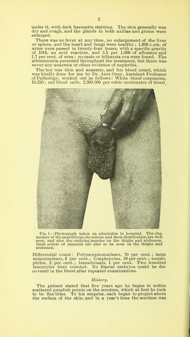
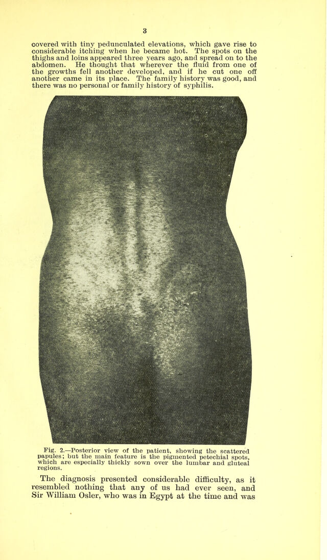
![Reprinted from the Bkitish Medical Journal, August 10th, 1912. PAPILLIFORM LESIONS (LYMPHANGIOMATA) OF THE SCROTUM, ASSOCIATED WITH MULTIPLE PETECHIAL SPOTS ON THE TRUNK AND LIMBS. Being a Paper read in the Section of Dermatology at the Annual Meeting of the British Medical Association,. Liverpool, 1912. By Frank Cole Madden, M.D.Melb., F.R.C.S.Eng., Professor of Surgery, Egyptian Government School of Medicine; Senior Surgeon Kasr-el-Ainy Hospital, Cairo. The subject of this peculiar condition was a young Egyptian fellah, Mohamed Amin, aged 23 years, from Minet-el-Gamh, Lower Egypt. He presented an extra- ordinary appearance, quite new to my experience, which I will endeavour to describe. The whole skin of the scrotum was thickly studded with small, succulent, pedunculated elevations, like thick-walled vesicles, purple or mulberry in colour, which were sprouting from the skin of the scrotum, the root of the penis, and the perineum as far back as the anterior margin of the anus. The elevations varied in size from a pin’s head to a pea, and presented all shades of colour from that of a morella cherry to a black grape. None of the skin of the scrotum was visible, but it appeared to be normal around the bases of the elevations, on separating them from each other with the finger. The root of the penis was sown with similar excrescences and a few were present on the body of the organ, but not on the glans. The perineum was in a similar condition, but more sparsely covered (Fig. 1). On cutting into the vesicles, which were quite painless and insensitiye, a dark haematin-stained fluid was discharged, but no fresh blood. Both thighs as far as the knees were covered with tiny dark purple spots, like subcuticular petechiae, and here and there raised papules of the same colour as the vesicles on the scrotum, which only rarely formed pedunculated elevations. The skin of the back, especially in its lower part and over the gluteal region, was more densely covered with these pigmented points, and a similar condition existed on the abdomen, and extended up as far as the costal margins (Fig. 2). There was an especially dark massing of these spots at the umbilicus, and a few scattered points also occurred on the inner sides of the elbows. These manifestations were apparently simple petechiae in the skin and Clinical Appearances, [422/12]](https://iiif.wellcomecollection.org/image/b22436868_0003.jp2/full/800%2C/0/default.jpg)