The pathology of tuberculous bone / by Cornelius Black.
- Black, Cornelius, 1822-1887.
- Date:
- 1859
Licence: Public Domain Mark
Credit: The pathology of tuberculous bone / by Cornelius Black. Source: Wellcome Collection.
Provider: This material has been provided by The Royal College of Surgeons of England. The original may be consulted at The Royal College of Surgeons of England.
8/40
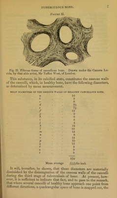
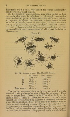
![diameter of which is often twice that of the osseous lamellaj inter- posed between adjacent cancelli. If a fine section of cancellous bone, from which the fat has been previously abstracted, be submitted to microscopic examination, numerous bodies, opaque in their appearance, will be seen in linear arrangement throughout the substance of each osseous lamella. These are the lacunae, which, in their figure, are either lenticular, oblong, irregularly ovate, or irregularly stellate. The first two forms represent the lacunae situated in the osseous lamellae between adja- cent cancelli, the mean measurements of which gave the following diameters:— Figure III. Fig. III.—Lacunae of bone—Magnified 500 diameters. A Long Diameter, inch. Short Diameter. 4000th inch. B 1 fh sB^rth „ C ffuVoth „ D 1 fh 5uWth » E 3shsth „ Mean average uiVgth „ The last two mentioned forms of lacunae are most frequently observed in the quadrangular islets of bone before named. These vary in length from 1-lOOOth to l-2000th of an inch, and in breadth from 1-1429th to l-2000th of an inch. ^ In the arrange- ment of the lacunas, their long diameter is invariably directed to- wards the adjacent cancellus. From their sides minute tubes or ])ores, termed canaliculi, proceed. These, in the series of lacunas lying nearest to a cancellus, communicate, on the one hand, with the latter, and, on the other, with the canaliculi of the lacunae next in order from the cancellus. They proceed nearly in straight lines. S](https://iiif.wellcomecollection.org/image/b22342588_0010.jp2/full/800%2C/0/default.jpg)

 No text description is available for this image
 No text description is available for this image
 No text description is available for this image