Bovine tuberculosis in man : an account of the pathology of suspected cases / by Charles Creighton.
- Charles Creighton
- Date:
- 1881
Licence: Public Domain Mark
Credit: Bovine tuberculosis in man : an account of the pathology of suspected cases / by Charles Creighton. Source: Wellcome Collection.
Provider: This material has been provided by The Royal College of Surgeons of England. The original may be consulted at The Royal College of Surgeons of England.
72/156 page 60
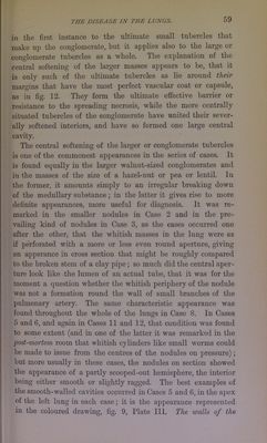
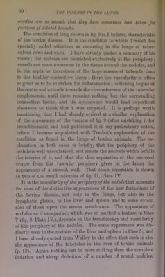
![«0 ntmitai are bo smooth that they hats sovietinus hrm taken for jMtrtions of dilated bronchi. Thu cotHlitioii of lunj( nIiowii in fig. 0 is, I bulievo, chanu-U*ristic of the bovine dimnise. It is tht* comlition to wliich Tmsbot bus 8|>eciully culled utteiilion us ocrurrinj' in the Imifjs of tulier- cnlons cows and oxen. I have alr«*U4ly quoted a suniinary of Iiis views; the nulules an* nourished exclusively at the jayriphery; vessels ore nion? nuniemus in the tissue around the nodules, and in the septa or interstices of the lai'|{e tnoiUM.*8 of tulH*n*le than in tljo healthy connwtive tissue; theh* the viiaoularity is ofteri soj^rent as to lie mistaken for influninmtion ; softening la^^ins at the (centre and extends tow4irds the cin'iimfen*nce of Uie tulx*rcle- conjjloniecate, until there remains nothing but tlie surrounding coiuaH'tive tissue, and its ap]M*urance would lead su]>eHicial ot»M*rverM to lliink that it was encysUsl. It is ]>erha|is worth mentioning, that I had nln*ady arrived at a similar ex]ilanation of the ap]M*araiux* of the vomica* of (ig. U (after mistaking it for bronchiectasis), and had publisluHl it in my preliminar}’ notice, liefon* 1 lx,*came aecpiainted with Traslnit's exphination of the condition ns found in tb* lungs of Ixivine animids. The ex- planation in lioth cases is briefly, that the |>eriphery of the iimlulo is well-vascularised, ajnl resists the necrt»sis which liefalls the interior of it, and that the clear Hcjianition of the necroscxl centre from the vascular periphery gives to the latU?r the np]>earance of a smooth wall. That clean sepamtion is shown in two of the small tulierclcs of tig. 11, Tlatc IV. It is the vascularity of the periphery of the nodule that accounts for most of the di.stinctive appearances of the new formations of the Iwvine disease, not only in the lungs, but also in the lymphatic glands, in the liver and sjileen, and to some extent also of those ujwn the semus membranes. The apiK^arancc of nodules as if encapsuled, which was so marked a feature in Case 7 (fig. 8, Plate IV.), depends on the translucency and vascularity of the iwrijihery of the nodules. The same apjicarance wjis dis- tinctly seen in the nodules of the liver nud spleen in Case 5; and I have already quoted from AValley to the effect that such is also tlie appearance of the tulierclcs in the liver of Ixivine animals (p. 12). Again, nothing can be more striking than the complete isolation and sharp definition of a number Df round nodules.](https://iiif.wellcomecollection.org/image/b2226758x_0074.jp2/full/800%2C/0/default.jpg)