How to use the ophthalmoscope : being elementary instructions in ophthalmoscopy, arranged for the use of students / by Edgar A. Browne.
- Browne, Edgar A. (Edgar Athelstane), 1841-1917.
- Date:
- 1883
Licence: Public Domain Mark
Credit: How to use the ophthalmoscope : being elementary instructions in ophthalmoscopy, arranged for the use of students / by Edgar A. Browne. Source: Wellcome Collection.
Provider: This material has been provided by The University of Leeds Library. The original may be consulted at The University of Leeds Library.
108/134 page 96
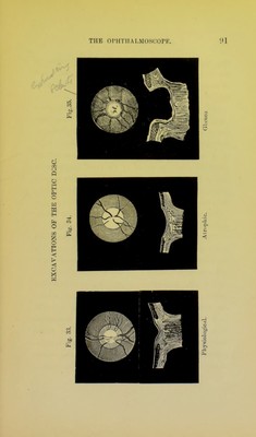
![( The constriction at tlie sclerotic ring impedes both the ingress and egress of blood. The veins are greatly enlarged, tortuous, and knotted. As they pass on to the surface of the disc, they are more or less covered by the exudation, sometimes completely; sometimes a bend will appear to dip beneath the surface and then reappear again, like portions of a line of railings cropping up here and there above the suiface of a snowdrift. {The arteries are reduced to mere threads, and are seen with difficulty, or not at all. A swollen disc is observed in tliree different conditions. (A.) When the predominant signs are those of vascular arrest. The ] )rqjection is very steep on one side, but slopes gradually down to the level of the disc on the other. The surface has a jnbssy look, and the margin of the disc is fringed with a fluffy grey border, which on close inspection can sometimes be made out as composed of innumerable small vessels and more or less semi-transparent effusion. The optic-nerve fibres are swollen and slightly opaque, so that they present a faint appearance of radia- tion. 'J'he colour varies from violet, reddish-grey, to a faint dusky gr^ scarcely tinged with colour. Numerous minute red spots are dotted over the](https://iiif.wellcomecollection.org/image/b2152046x_0108.jp2/full/800%2C/0/default.jpg)


 No text description is available for this image
 No text description is available for this image
 No text description is available for this image