A report of microscopical and physiological researches into the nature of the agent or agents producing cholera / by T.R. Lewis and D.D. Cunningham.
- Timothy Richards Lewis
- Date:
- 1872
Licence: Public Domain Mark
Credit: A report of microscopical and physiological researches into the nature of the agent or agents producing cholera / by T.R. Lewis and D.D. Cunningham. Source: Wellcome Collection.
53/126 page 43
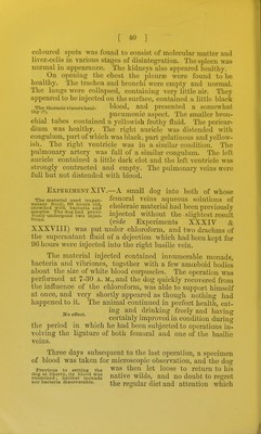
![[ ^-13 ] food, and was slightly congested. Towards its pyloric ex- tremity there was a very hard fihroid tumour apparent- ly of a schirrous nature. The liver appeared healthy; there were numerous disfomata in the bile ducts.* There were no traces of embolism throughout its substance. The ffall-bladder contained bile. The spleen and kidneys were healthy. On opening the thorax, the pleural cavities were lis. C XSOO A. Fat.Si'/^c D. X500 Oral sucker. CEsophagus. ^ Termination of genital tube \ with ova escaping. Ventral sucker, protruded. Alimentary canal, right side. Uterine tuhe filled with ova. Hairs, still adherent. Vitellogeno glands, right side. Right vitellogene duct. Ovary, Eiffht testicle. Termination of right alimentary tube. Pulsotile vesicle, near termina- tion of water-vascular system. X.7S * Fig. 3, DiSTOMA POUND IN THE BIIE DUCTS OF DOGS. A. —The parasite figured natural size, B. —Ditto magnified 15 diameters. C. —Minute hairs covering the entire body when fresh and before being manipulated, magnified 300 diameters, D. —Ova squeezed out of the uterine tube, magnified 300 diameters. This distoma is not infrequently met with in the bile ducts of dogs in this country. With the limited supply of literature on this subject within our reach, we have, however, not been able to refer it to any described species, and have therefore introduced a wood-cut showing its size, form and minute anatomy, together with those of the ova. It appears to us to be very closely allied to the species discovered by Dr. Cobbold in the liver of the American red fox, and described and figured by him in his valuable work on Entozoa; indeed if, on re-examination it be found that that parasite has been, inadvertently, drawn by Dr. Cobbold as seen from the back—a mistake into which we ourselves fell, when the first specimen was sketched—this may turn out to be identical with the species described by this Author under the name of Distoma Conjuncium. We strongly suspect this to be the case.](https://iiif.wellcomecollection.org/image/b20392023_0053.jp2/full/800%2C/0/default.jpg)