On the compound vision and the morphology of the eye in insects / by B. Thompson Lowne.
- Lowne, B. Thompson (Benjamin Thompson), 1839-1925.
- Date:
- 1884
Licence: Public Domain Mark
Credit: On the compound vision and the morphology of the eye in insects / by B. Thompson Lowne. Source: Wellcome Collection.
11/172 page 391
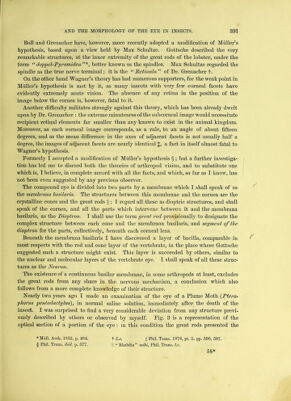
![Boll and Grenadier have, however, more recently adopted a modification of Muller’s hypothesis, based upon a view held by Max Schultze. Gottsclie described the very remarkable structures, at the inner extremity of the great rods of the lobster, under the term “ doppel-Pyramiden better known as the spindles. Max Schultze regarded the spindle as the true nerve terminal; it is the “ Petinula ” of Dr. Grenacher f. On the other hand Wagner’s theory has had numerous supporters, for the weak point in Muller’s hypothesis is met by it, as many insects with very few corneal facets have evidently extremely acute vision. The absence of any retina in the position of the image below the cornea is, however, fatal to it. Another difficulty militates strongly against this theory, which has been already dwelt upon by Dr. Grenacher : the extreme minuteness of the subcorneal image would necessitate recipient retinal elements far smaller than any known to exist in the animal kingdom. Moreover, as each corneal image corresponds, as a rule, to an angle of about fifteen degrees, and as the mean difference in the axes of adjacent facets is not usually half a degree, the images of adjacent facets are nearly identical $, a fact in itself almost fatal to Wagner’s hypothesis. Formerly I accepted a modification of Muller’s hypothesis § ; but a further investiga- tion has led me to discard both the theories of arthropod vision, and to substitute one which is, I believe, in complete accord with all the facts, and which, so far as I know, has not been even suggested by any previous observer. The compound eye is divided into two parts by a membrane which I shall speak of as the membrana basilaris. The structures between this membrane and the cornea are the crystalline cones and the great rods |; I regard all these as dioptric structures, and shall speak of the cornea, and all the parts which intervene between it and the membrana basilaris, as the Dioptron. I shall use the term great rod provisionally to designate the complex structure between each cone and the membrana basilaris, and segment of the dioptron for the parts, collectively, beneath each corneal lens. Beneath the membrana basilaris I have discovered a layer of bacilla, comparable in most respects with the rod and cone layer of the vertebrate, in the place where Gottsche suggested such a structure might exist. This layer is succeeded by others, similar to the nuclear and molecular layers of the vertebrate eye. I shall speak of all these struc- tures as the Neuron. The existence of a continuous basilar membrane, in some arthropods at least, excludes the great rods from any share in the nervous mechanism, a conclusion which also follows from a more complete knowledge of their structure. Nearly two years ago I made an examination of the eye of a Plume Moth (Ptero- yhorus pentadactylus), in normal saline solution, immediately after the death of the insect. I was surprised to find a very considerable deviation from any structure previ- ously described by others or observed by myself. Pig. 3 is a representation of the optical section of a portion of the eye : in this condition the great rods presented the * Miill. Arch. 1852, p. 484. f Lx. + Phil. Trans. 1878, pt. ii. pp. 596, 597. § Phil. Trans, ibid. p. 577. |] “ Rhabdia ” mihi, Phil. Trans, lx. 58*](https://iiif.wellcomecollection.org/image/b28066595_0011.jp2/full/800%2C/0/default.jpg)