On the development of the blood and blood-vessels, being the prize essay for 1854 of the Edinburgh Harveian Society / by James Drummond, M.D.
- Drummond, James
- Date:
- [1854]
Licence: Public Domain Mark
Credit: On the development of the blood and blood-vessels, being the prize essay for 1854 of the Edinburgh Harveian Society / by James Drummond, M.D. Source: Wellcome Collection.
Provider: This material has been provided by The University of Glasgow Library. The original may be consulted at The University of Glasgow Library.
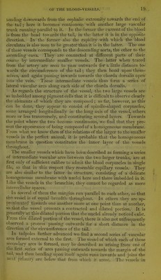
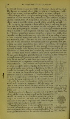
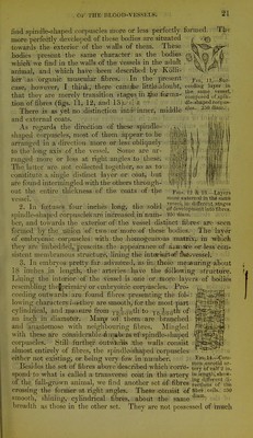
21/24 page 21
![Tlie Fig, ]],—Suc- ceeding layer in the same vessel, ■ composed of.spm- dle-shaped corpus- cles,. 'iCiO diam.i, find spindle-shaped corpuscles more or less perfectly formed, more perfectly developed of these bodies are situated towards the exterior of the walls of them. These bodies present the same character as the bodies which we find in the walls of the vessels in the adult animal, and which have been described by Kolli- kei' .as organic muscular fibres. In the present case, however, I think,; there can?.{be little adoubt, that they are merely transition stages, in .tiie forma- tion of fibres (figs. 11, 12, and 13). '. - ! fc There is as yet no distinction into-^'inner, middle and external coats. As regards the direction of these spindle- shaped coi'puscles, most of them appear to be arranged in a direction, niore or less obliquely to the long axis of the vessel. Some are ar- ranged more or less at right angles to these. The latter are not collected together, so as to constitute a single distinct layer or coat, but are found intermingled with the others through- out the entire thickness of the coats of the vessel. 2. In foetuses four inches long, the solid spindle-shaped corpuscles are increased.in num- 250 dinm'. ber, and towards the exterior o£'the ivessel distinct fibres'are seen formed by the union of two; or more of these bodies. ■ The layer of embryonic corpuscles with the homo^ijeueous mati;ixj. in which tliey are imbedded, presents: the appearance of awmoi'e or less con- sistent membranous structure, lining the inteirititicvf {liiavvesseli' * '^'j 3. In embryos pretty far advajlced, as in/ those measuring'about 18 inches in length, the arteries- ;have the ^£61 lowing structure. Lining the interior of 'the vessel is .lone or taore layers of bodies resembling tliejprimary or embrydnic coi'puscles. Pro- ceeding outwards are found fibres presenting the fol'- lowing characters.3r*they are smooth,'for the most part cylindrical, and mea-rare from ^-2.^-(Y;(^th'to xo.oou^'^ an inch in diameter. Maa\y-.of them::are branched II m Figs, f 2 '1.3 —L&'i'ers Ttiorp extci-rial in thi same vessel-, in difj'ereut,. stages of devcloptaenit inio fibres. Mingled and anastomose with neighbouring fibres with these are cdnsiderable attuiabeiiStff!spindle-shaped corpuscles. Still furthelr outuiirils Itlie walls consist almost entirely of fibres, the spindle-shaped corpuscles either not existing, or being very few in number. ; Besides the set of fibres above described which corre- spond to what is called a transverse coat in the artery of the full-grown animal, we find another set of fibi'es (•ro.ssing the former at right angles. These >c6nsist 'smooth, shining, cylindrical fibres, about breadth as those in the other set. They are not possessed of !much Fio, 14^.—Com- mon carotid ar- tery of calf 2 in. in leng(li, .show- ing djtttH'cnt di- rections of die of librb cella. 250 .1 diam.. . ■ the same - ■ i. • • ■](https://iiif.wellcomecollection.org/image/b21477644_0021.jp2/full/800%2C/0/default.jpg)