Guide to the examination of urine : with special reference to the diseases of the urinary apparatus / by K.B. Hoffman ... and R. Ultzmann.
- Karl Berthold Hofmann
- Date:
- 1879
Licence: Public Domain Mark
Credit: Guide to the examination of urine : with special reference to the diseases of the urinary apparatus / by K.B. Hoffman ... and R. Ultzmann. Source: Wellcome Collection.
Provider: This material has been provided by the Francis A. Countway Library of Medicine, through the Medical Heritage Library. The original may be consulted at the Francis A. Countway Library of Medicine, Harvard Medical School.
208/218 page 192
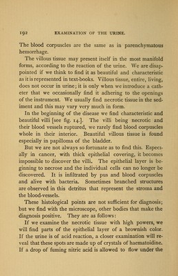
![The blood corpuscles are the same as in parenchymatous hemorrhage. The villous tissue may present itself in the most manifold forms, according to the reaction of the urine. We are disap- pointed if we think to find it as beautiful and characteristic as it is represented in text-books. Villous tissue, entire, living, does not occur in urine; it is only when we introduce a cath- eter that we occasionally find it adhering to the openings of the instrument. We usually find necrotic tissue in the sed- iment and this may vary very much in form. In the beginning of the disease we find characteristic and beautiful villi [see fig. 14.]. The villi being necrotic and their blood vessels ruptured, we rarely find blood corpuscles whole in their interior. Beautiful villous tissue is found especially in papilloma of the bladder. But we are not always so fortunate as to find this. Especi- ally in cancer, with thick epithelial covering, it becomes impossible to discover the villi. The epithelial layer is be- ginning to necrose and the individual cells can no longer be discovered. It is infiltrated by pus and blood corpuscles and alive with bacteria. Sometimes branched structures are observed in this detritus that represent the stroma and the blood-vessels. These histological points are not sufficient for diagnosis; but we find with the microscope, other bodies that make the diagnosis positive. They are as follows: If we examine the necrotic tissue with high powers, we will find parts of the epithelial layer of a brownish color. If the urine is of acid reaction, a closer examination will re- veal that these spots are made up of crystals of haematoidine. If a drop of fuming nitric acid is allowed to flow under the](https://iiif.wellcomecollection.org/image/b21059226_0208.jp2/full/800%2C/0/default.jpg)