Exhibition and description of the skull of a microcephalic Hindu / by R.W. Reid.
- Reid, Robert William, 1851-1939.
- Date:
- 1894
Licence: Public Domain Mark
Credit: Exhibition and description of the skull of a microcephalic Hindu / by R.W. Reid. Source: Wellcome Collection.
Provider: This material has been provided by The Royal College of Surgeons of England. The original may be consulted at The Royal College of Surgeons of England.
5/10 (page 107)
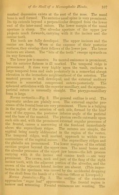
![marked depression exists at the root of the nose. The nasal bone is well formed. The anterior nasal spine is very prominent. Its tip extends beyond a perpendicular dropped from the lower end of the inter-nasal suture. The lower margin of the nasal aperture is sliarp. The alveolar aperture of the upper jaw projects much forwards, carrying with it the incisor and the canine teeth. The teeth are fully developed. The upper incisors and the canine are large. Worn at the expense of their posterior surfaces, they overlap their fellows of the lower jaw. The lower incisors are absent. The “bite of the teeth” sweeps upwards on its way forwards. The lower jaw is massive. Its mental eminence is prominent, but its anterior flatness is ill marked. The temporal ridge is pronounced. It rises very highly upon the vault, encloses a large temporal fossa and ends posteriorly in a somewhat abrupt elevation in the immediate neighbourhood of the asterion. The mastoid process is well developed, and the external auditory meatus is somewhat compressed antero-posteriorly. The sphenoid articulates with the superior maxillary, and the squamo- parietal suture is unusually straight. The pterygo-maxillary fossa is large. Norma veodiGaUs.—Fig. 2. The general ontline is oval. The zygomatic arches are plainly seen. The external angular pro- cesses of the frontal bone are very prominent. There is a bulging- in the region of the asterion of each side, formed by the hind root of the zygoma, the posterior inferior angle of the parietal and the base of the mastoid. The pterion swells outwards upon each side and, with the prominent external angular processes of the frontal, causes a narrowing of the forehead immediately above the supraciliary ridges. The sutures are simple, the s^ittal being nearly obliterated in the region of the vertex. 1 he temporal ridges, well marked, approach to within two centimetres of the sagittal suture. The supraciliary ridges and the glabella are pronounced. The lower margins of the orbital cavities project beyond the upper ones. The nasal bones and the alveolar processes of the upper jaw bones—the left process carrying with it the incisor and canine teeth — are very prominent. The crown, neck and part of the fang of the right incisor tooth, with the adjacent portion of the alveolus, and the crown ot the adjacent lateral incisor are absent. (This imper- tection in the specimen was caused by the accidental dropiiing of the skull from the hands of a Customs Officer at Liverpool.) oima fiontalis. Fig. 3. The outline of the cranium approac les sugar loaf ap])earaiices. The frontal region is narrow and retreating. Frontal eminences are wanting. The](https://iiif.wellcomecollection.org/image/b22381028_0007.jp2/full/800%2C/0/default.jpg)