A case of primary tumor of the optic nerve / by F. Buller.
- Buller, Frank, 1844-
- Date:
- 1899
Licence: Public Domain Mark
Credit: A case of primary tumor of the optic nerve / by F. Buller. Source: Wellcome Collection.
Provider: This material has been provided by The Royal College of Surgeons of England. The original may be consulted at The Royal College of Surgeons of England.
6/18 page 4
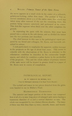
![the nerve appears as a small circle in the center of a yellowish, gelatinous-looking-, lathei firm mass, ihe portion at first re- moved constitutes about 2/3 of the entire mass, that which was taken away after removal of the eye the remaining third, this portion being concave anteriorly and perforated at its center. This disk-like segment when fitted over the other completes the growth. In separating the parts with the scissors, they must have passed into a sulcus in the soft tissues, and so divided the tumor into the two portions just described. The chief interest in this case is the pathological report pre- pared by my friend and colleague, Dr. Byers, and herewith sub- mitted in extenso. I wish particularly to emphasize the apparent, sudden increase in the proptosis at the age of about four years. This seems to coincide with the observations of Dr. Byers in reference to the oedematous condition he describes in the extraocular lymph- spaces, and which he considers a prime factor in the production of the proptosis. This and the whole subject of primary tumors of the optic nerve will be treated in greater detail in a paper of Dr. Byers now in course of compilation. PATHOLOGICAL REPORT. By W. GORDON M. BYERS, M.D. [From the Pathological Laboratory of the Royal Victoria Hospital.] The eyeball and tumor in two pieces detached from the globe were handed to me in Muller's Fluid. Macroscopical Examination. The anterior and larger portion of the tumor was composed first of all of a 15 mm. long piece of enlarged optic nerve having a transverse diameter of 12 and a vertical diameter 10 mm. The whole was encapsuled by two distinct fibrous sheaths. The inner of these was thin (less than 0.5 mm.), smooth, dense, and loosely](https://iiif.wellcomecollection.org/image/b22368061_0008.jp2/full/800%2C/0/default.jpg)