Twelve lectures on comparative embryology : delivered before the Lowell Institute, in Boston, December and January, 1848-9 / by Louis Agassiz ... Phonographic report, by James W. Stone ... Originally reported and published in the Boston Daily Evening Traveller.
- Louis Agassiz
- Date:
- 1849
Licence: Public Domain Mark
Credit: Twelve lectures on comparative embryology : delivered before the Lowell Institute, in Boston, December and January, 1848-9 / by Louis Agassiz ... Phonographic report, by James W. Stone ... Originally reported and published in the Boston Daily Evening Traveller. Source: Wellcome Collection.
19/116 (page 13)
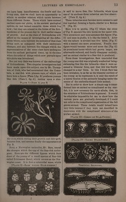
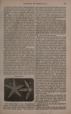
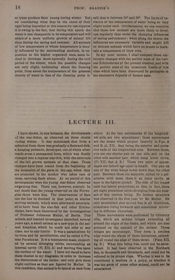
![ces have been foundbetween the fossils and the living ones, that we shall have an opportunity to allude to another relation which exists between these different forms. Those which have existed earliest upon cur globe, in the ancient geological epdéchs, do not indeed resemble those which live now ; but they are related to the forms of the Ech- inoderms of the present day in their earlier stages of growth. And so the class of Echinoderms will afford us the means of investigating all the differ- ences which exist between the animals of that class living now, as compared with their embryonic changes, and also between the changes which: the representatives of the same class have undergone from the earliest geological times, up to the time when theorder of things which now prevails upon this globe was introduced. But yet very little was known of the embryology of Echinoderms. Two singular investigations had been made upon this subject, one by Mr. Thomp- son, of Cork, who had ascertained that the Coma- tula, a star-fish with pinnate rays, of which you have here a figure [Plate I,fig. B] produces youngs like this [Plate I, fig. A], resting upon a slen- PratEeE I—Fres A ann B. der stem,which during their growth cast this stem, become free, and assume finally the appearance of Fig. B. Next, a Norwegian naturalist, Mr. Sars, traced the changes which the egg of the Star-fish under- goes. Here are the different figures which Sars drew of the young of a small species of Star-fish called Echinaster Sarsii, which occurs on the Nor- wegian coast. It is first a spheroidal mass, which [PLate Ii—Sars’ youne STAR-FISHES.] 7 E ttaaes s * EMBRYOLOGY. 13 is said to move free, like Infusoria, when upon one of its surfaces three tubercles are first observ- ed. [Plate IL, fig A]. These tubercles soon become more extensive and run together. forming a figure, similar to a Roman T. [Fig. B]. Here itis in profile, [Fig.C] where the cross of Fig. B. appears like two horns on the upper side. This prominent part next assumes this figure [Fig. D] and seen in profile, it is like the letter E, After this the sphere is divided into five lobes, [Fig. F] with a central one more prominent. Finally, that figure would become more and more flat [Fig. G] its prominent horns which had grown larger, are afterwards reduced, and finally disappear entirely, and an animal similar to a Star-fish is produced. From these investigations, Sars concluded that the young star-fish was originally aspherical being» swimming free like the infusoria—that it soon as- sumed a bilateral form, and that this was finally changed to astar form. In this I think Sars has been mistaken, in as far as the bilateral outlines of the young as he represents it, is only the result of a lateral flexion of the peduncle hanging under the centre of the umbrella-shaped little animal. But in order to show how a simple egg is trans- formed into an animal so complicated as the star- fish, it is now necessary for meto allude, first,. to the structure of Echinoderms in general. It would be otherwise impossible for me to show how the various parts are gradually developed, if I could not refer to the complicated organization of the full grown animal. These Getails would indeed have](https://iiif.wellcomecollection.org/image/b33278982_0019.jp2/full/800%2C/0/default.jpg)