Twelve lectures on comparative embryology : delivered before the Lowell Institute, in Boston, December and January, 1848-9 / by Louis Agassiz ... Phonographic report, by James W. Stone ... Originally reported and published in the Boston Daily Evening Traveller.
- Louis Agassiz
- Date:
- 1849
Licence: Public Domain Mark
Credit: Twelve lectures on comparative embryology : delivered before the Lowell Institute, in Boston, December and January, 1848-9 / by Louis Agassiz ... Phonographic report, by James W. Stone ... Originally reported and published in the Boston Daily Evening Traveller. Source: Wellcome Collection.
21/116 (page 15)
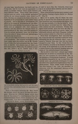
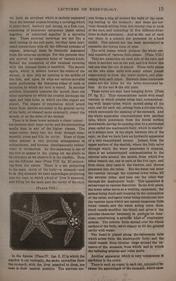
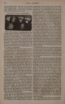
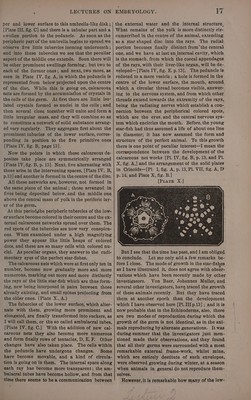
![we have an envelope which is entirely separated from the internal organs,forming a covering,which is either hard, leathery and strong, or a firm coat, “consisting of numerous calcareous plates united together, or connected together in a movable way. These external coverings are not like a shell resting on the soft parts, but they form intri- cated connections with all the different systems of organs, altheugh these be distinctly separated from the external envelope. For this purpose they are pierced by numerous holes of various kinds. indeed the conrexion of this external covering with the internal frame is manifold. The mouth again, which is always toward the centre of the animal, is also only an opening in the middle of the disk, and upon its edge are various movabie parts performing the functions either of teeth or tentacles, by which the food is seized. In another position, frequently opposite the mouth, there are other apertures by which the ovaries discharge the eggs, and little holes in which eye-like organs are placed. The organs of respiration which“ admit water from outside are either in the general cavity ‘of the body, or situated more externally, round the mouth, or on the sides of the animal. There is in these lower animals a closer connex- ion between their inner cavity and the surrounding media than in any of the higher classes. The water rushes freely into the body through innu- merabie pores and fills its cavity. Some of these tubes assume a very peculiar arrangement in echinoderms, and become simultaneously subser- vient to locomotion. As this apparatus is one of the first to appearin the young, let me allude to its structure as we observe it in the starfish. Here are the diferent rays ]Plate VIII fig. B] project- ing from the centre. starfish is cut vertically, the sacks extending from the stomach, with the liver attached to them, are seen in their natural position. The nervous sys- 15 tem forms a ring all around the walls of the open- ing leading to the stomach; and there are ner- vous threads arising from this central ring to each of the rays, and extending in five different direc- tions to their extremity. And atthe end of each ray there is a colored dot protected by a hard shield. This colored dot has been ascertained to resemble the lowest form of eyes. The solid frame which protects the whole ani- mal consists of various little plates. {Plate VIII.] They are numerous on each side of the rays, and there is another one at the end, and itis below this last one that the eye is placed. Those solid plates on the two sides unite with many others placed transversely to form the lower surface, and alter- nating with each other. Between these transverse plates are the holes for the tubes mentioned be- fore. At the end is the odd plate. These tubes are seen here hanging down [Plate IV. fig. H.]. They communicate inside with small vesicles, to which minute tubes lead, communicat- ing with larger tubes, which extend along all the rays, one for each ray, arising from a circular tube, which surrounds the opening of the stomach. And the whole apparatus communicates with another tube, which penetrates from the dorsal surface downwards, having its opening shut by a perforated plate called the madreporic body, which in starfish- es is always seen in the angle between two of the rays; so that we have here an hydraulic apparatus of avery complicated nature. Indeed, from the upper surface of the starfish, where the littie seive through which the water penetrates is situated, there is an uninterrupted communication to the circular tube around the mouth, from which five tubes branch out, one to each of the five rays; and from these, they open to the vesicles, and thence penetrate into the tubes. But the water can enter the vesicles through the external-lower tubes, fill the eircular tubes, and* pass out the other way through the madreporic body. This apparatus is subservient to various functions. In the first place, the lower tubes serve as a walking apparatus; the animal being fixed and creeping by the contraction of the tubes, and again water being introduced into the vesicles upon which are spread numerous little blood vessels, and the water acting upon these blood vesseis modifies the blood, and gives it the peculiar character necessary to perform its func- tions, constituting a peculiar kind of respi atory system. The minute holes spread over the whole surface of the body, serve simply to fill the general cavity with water. The heart is placed along the calcareous tube which arises from the madreporic body, and the bleod vessels form circular rings around the en: trance of the stomach, from which and to which the radiating arteries and veins move. - Another apparatus which is very volaminous in starfishes is the ovary. There is such an organ in each ray, concealed be- tween the appendages of the stomach, which open](https://iiif.wellcomecollection.org/image/b33278982_0021.jp2/full/800%2C/0/default.jpg)