Twelve lectures on comparative embryology : delivered before the Lowell Institute, in Boston, December and January, 1848-9 / by Louis Agassiz ... Phonographic report, by James W. Stone ... Originally reported and published in the Boston Daily Evening Traveller.
- Louis Agassiz
- Date:
- 1849
Licence: Public Domain Mark
Credit: Twelve lectures on comparative embryology : delivered before the Lowell Institute, in Boston, December and January, 1848-9 / by Louis Agassiz ... Phonographic report, by James W. Stone ... Originally reported and published in the Boston Daily Evening Traveller. Source: Wellcome Collection.
22/116 (page 16)
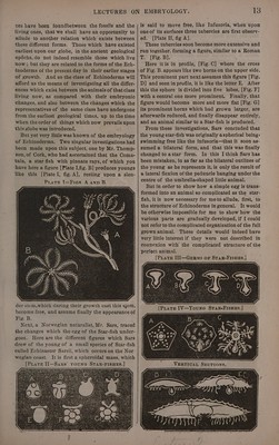
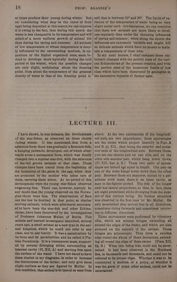
![upon the upper surface with little holes through which the eggs escape. The ovary itself is a gra- nular organ, of which several figures in various stages of developement are here seen. “PLateE [X—Ovaries oF StTar-F1suH.] Such is the structuré of the Echinoderms, the stomach forming a simple cavity, without any oth- er outlet except the mouth. In some the alimen- tary tube is more complicated. Inthe Echini or Sea-Urchins [Plate VI] there is an alimentary tube forming several evolutions, and opening upwards. The ovaries form more peculiar masses than in the star-fishes. The mouth is also protected in most Echini by a complicated set of jaws and teeth. In Holothuris the whole system of organs assumes & more bipartite arrangement. In the process which gives rise to the formation of new individuals, the first step consists in the ac- cumulation of more or less consistent matter of a somewhat opaque or yellowish appearance, and of & granulated texture which divides soon into small spherical masses. This takes place in the ovary.— This mass, at first homogeneous, assumes soon the aspect of little bunches, which soon grow more and more isolated, and then assume around them a pe- culiar membrane, and there areeggs. Eggs in their simplest condition are microscopical spheres of a homogeneous mass, called yolk, and surrounded by a simple membrane, called the yolk membrane. (Plate IX C.) However different in its aspect in different animals, this mass is called yolk, through- out the animal kingdom, from the fact that this name has been applied to the part which corres- ponds to this structure in the hen’s egg. The primitive egg is always microscopical, and its contents homogeneous ; but this substance soon becomes granular. It is so small as to escape the observation 6f the naked eye. And there is anoth- er little sphere formed within, which is called the germinative vesicle, containing another little ves- cicle, which is called the germinative dot. [Plate IX fig.D.] Undera powerful microscope the gran- ules of the yolk itself appear also like little cells.— There are little spherical masses, and they contain even in their turn other little dots. Plate IX shows the various degrees of the growth of such eggs, of which there are more or less de- veloped ones in the same ovary ; assuming first their regular form [Fig. A], and then a transparent space appearing in the interior [Fig.B] ; next the germinative vescicle becomes more distinct [Fig. C], and [Fig. D] the germinative dot is now dis- tinctly seen. The whole mass of yolk, which has grown considerably; consists here of cells, which have been formed by the expansion of its granules. ? Through this growth of cells within cells, and of granules growing into cells, there is finally a germ formed. That which we call yolk in the beginning, is finally a spherical germ, which will escape from its envelope. We have here [Fig. E] the ovary of a Star-fish, from which some germs have escaped, and here is the figure of such a germ already hatched, highly magnified [Fig. F]. The ovary of sea urchins, have all the same structure, and vary only in their size and proportions. Nowa curious observation which I have had an opportuni- ty to make, is, that the eggs after they are laid are taken up by the star-fish, and kept between its tubes, below the mouth. The star-tish bends itself around them, surrounds the eggs with its suckers, and moves about with them. When the eggs had been removed to some distance from the animal, it went towards them and took them up again, and moved off with them. showing that these animals, so low in structure, and apparently deprived of all. instinct, really have so much instinct as to watch over their young. Now these eggs which are thus kept there, and protected by the mother, will escape. These germs I have been able to trace from the lowest possible condition, where they resemble ovarian eggs. At no epoch didI see this new born animal living free, and swimming like Infusoria, as is said to be the case by Sars. Soon, however, the external crust of the germ becomes more transparent, consisting of somewhat looser and larger granules, and the internal mass assumes a color a little darker, so that two layers are distinct, between which there is another one, which becomes also gradually more and more dis- tinct. On one side of the germ there is now a pro- tuberance forming, and the prominent portion separates more and more from the spherical mass, [Plate IX, F] the difference in substance of its layers growing more and more distinct. The promi- nent portion, which is the lower part of the little animal, becomes more and more elongated and as- sumes more and more the form of a peduncle. Often there are several grouped together, and at- tached by this appendage to the empty egg cases}; they would even-form bunches remaining thus attached till they are far advanced in their growth. At this period, however, there is not yet any or- gan formed as you will notice on comparing Fig. F of Plate IX. with those of PlateIV.p.33. Only changes of substance have taken place. But now we begin to see little swellings in five points om the sides; the spherical portion of the germ has also grown considerably, and has been flattened by lateral dilatation. The little animal has grown to a more hemis- pherical shape; and from that time there is an up-](https://iiif.wellcomecollection.org/image/b33278982_0022.jp2/full/800%2C/0/default.jpg)