Twelve lectures on comparative embryology : delivered before the Lowell Institute, in Boston, December and January, 1848-9 / by Louis Agassiz ... Phonographic report, by James W. Stone ... Originally reported and published in the Boston Daily Evening Traveller.
- Louis Agassiz
- Date:
- 1849
Licence: Public Domain Mark
Credit: Twelve lectures on comparative embryology : delivered before the Lowell Institute, in Boston, December and January, 1848-9 / by Louis Agassiz ... Phonographic report, by James W. Stone ... Originally reported and published in the Boston Daily Evening Traveller. Source: Wellcome Collection.
23/116 (page 17)
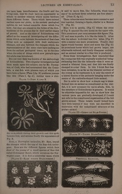
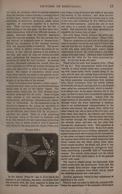
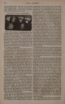
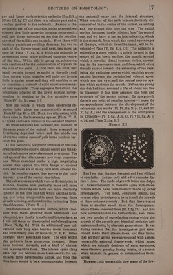
![per and lower surface to this umbrella-like disk ; [Plate ILI, fig. C] and there is a tubular part and a swollen portion to the peduncle. As soon as the peripheric part of the umbrella begins to spread,we observe five little tubercles forming underneath ; and into these tubercles we see that the peculiar aspect of the middle one extends. Soon there will be other prominent swellings forming; but two to each of the former ones; and next, two more, as seen in Plate IV, fig. A, in which the peduncle is represented from below projected upon the centre of the disc. While this is going on, calcareous nets are formed by the accumulation of crystals in the cells of the germ. At first there are little iso- lated crystals formed as nuclei in the cells ; and then several close together will unite and form a little irregular mass, and they will combine so as to constitute a network of solid substance arrang- ed very regularly. They aggregate first about the prominent tubucles of the lower surface, corres- ponding in. position to the five primitive ones [Plate IV, fig. B, page 13]. Now the points in which these calcareous de- posites take place are symmetrically arranged [Plate IV, fig. B, p. 13]. Next, five alternating with these arise in the intervening spaces, [Plate IV, B, p.13] and another is formed in the centre of the disc. All these networks are, however, not formed in the same plane of the animal; those arranged in fives being deposited below, and the.middle one above the central mass of yolk in the periferic lay- er of the germ. At this period,the peripheric tubercles of the low- er surface become colored in their centre and the ex- ternal calcareous networks spread over them. The red spots of the tubercles are now very conspicu- ous. When examined under a high magnifying power they appear like little heaps of colored dots, and these are so many cells with colored nu- clei. As peculiar organs, they answer to the rudi- mentary eyes of the perfect star-fishes. The calcareous nets which were at first only ten in number, become now gradually more and more numerous, marking out more and more distinctly the rays of the little star-fish which are thus form- ing, new being interposed in pairs between those already existing, and small spines projecting from the older ones. (Plate X., A.) The fubercles of the lower surface, which alter- nate with them, growing more prominent and elongated, are finally transformed into suckers, as I will call them, or the so called ambulacral tubes, [Plate IV, fig.C.] With the addition of new cal- careous nets they also become more numerous and form finally rows of tentacles, D, E, F. Other changes have also taken place. The cella within the peduncle have undergone changes. Some haye become movable, and a kind of circula- tion is going on in them. The internal space along each ray has become more transparent; the am- bulacral tubes have become hollow, and from that time there seems to be a communication between the external water and the internal structure, What remains of the volk is more distinctly cir- cumscribed in the centre of the animal, extending as a star-shaped disc into the rays. The radial portion becomes finally distinct from*the central one, and we have at last an internal cavity, which is the stomach, from which the coecal appendages of the rays, with their liver-like organ, will be de~ veloped—[Plate IV, fig. E. p.13]. The peduncle is reduced to a mere vesicle; a hole is formed in the centre of the lower surface, the mouth, around which a circular thread becomes visible, answer- ing to the nervous system, and from which other threads extend towards the extremity of the rays, being the radiating nerves which establish a con- nection between the peripherical colored spots, which are the eyes, and the central nervous sys- tem which encircles the mouth. Before, the young star-fish had thus assumed a life of about one line in diameter; it has now assumed the form and stracture of the perfect animal. To this growth there is one point of peculiar interest—I mean the correspondence between the development of the calcareous net works [Pi. IV, fig. B, p. 13, and Pl. X, fig. A,] and the arrangement of the solid plates in Crinoids—[PI. I, fig. A, p. 13, Pl. VII, fig. A, D p. 14, and Plate X, fig. B.] [PuatTEe X.] But I see that the time has past, and lam obliged to conclude. Let me only add a few remarks be- fore I close. The mode of growth in the star-fishes as I have illustrated it, does not agree with obser- vations which have been recently made by other investigators. Von Baer, Johannes Muller, and several other investigators, have traced the growth of these animals recently. But they have traced them at another epoch than the development which [ have observed here [PI. III p.13] ; and it is now probable that in the Echinoderms, also, there are two modes of reproduction during which the growth of the germ is not identical, as in the ani- mals reproducing by alternate generations. It was during summer that the investigators just men- tioned made their observations, and they found that all their germs were surrounded with a most remarkable external frame-work, whilst mine, which are entirely destitute of such envelopes, were observed growing during winter, at a season when animals in general do not reproduce them- selves. However, it is remarkable how many of the low-](https://iiif.wellcomecollection.org/image/b33278982_0023.jp2/full/800%2C/0/default.jpg)