Twelve lectures on comparative embryology : delivered before the Lowell Institute, in Boston, December and January, 1848-9 / by Louis Agassiz ... Phonographic report, by James W. Stone ... Originally reported and published in the Boston Daily Evening Traveller.
- Louis Agassiz
- Date:
- 1849
Licence: Public Domain Mark
Credit: Twelve lectures on comparative embryology : delivered before the Lowell Institute, in Boston, December and January, 1848-9 / by Louis Agassiz ... Phonographic report, by James W. Stone ... Originally reported and published in the Boston Daily Evening Traveller. Source: Wellcome Collection.
45/116 (page 39)
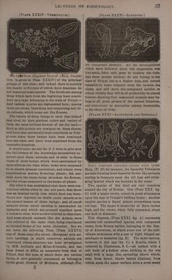
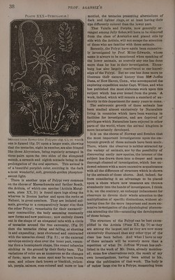
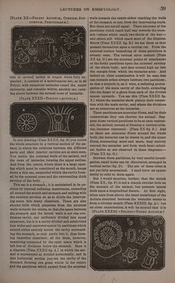
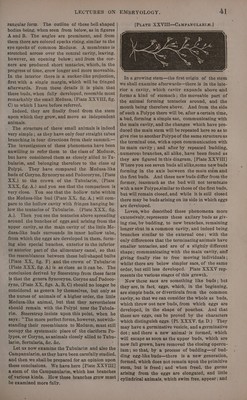
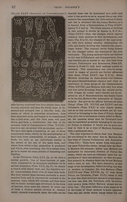
![one to several inches in length when fully ex- panded ; it consists of a membranous sac, as in all Polypi, with numerous tentacies round the upper extremity, and contains within, another sac, open- ing above between the several rows of tentacles. [PLATE XX XII—PoLyPI—ActTin1I#. | In this drawing {Piate XXXII, fig. B) you notice the whole structure in a vertical section of the an- imal, in which the relations between the different parts and their interior cavities are at once seen. You notice the external walls of the animal, and the rows of tentacles forming the upper outline. And from the centre, where there is a large open- ing which must be considered as the mouth, hangs down a thin sac, suspended within the cavity form- ed by the external arms and the surrounding thick envelop of the body. , This sac is a stomach; it is maintained in its po- sition by internal radiating membranes, extending all around the mouth and stomach and uniting with the external envelop so as to divide the interven- ing space into many chambers. There are also shorter folds which penetrate from the external walls towards the centre; so that the space between the stomach and the lateral walls is not one con- tinuous cavity, nor uniformly divided into equal chambers, but it is a cavity divided and subdivided into wider and narrower spaces by partitions which extend either entirely across the cavity surround- ing the stomach, or only partly into it, thus form- ing imperfect chambers; all the them, however, remaining connected by the open space which is left free of divisions below the stomach. Here is a diagram [Plate XXXII fig. A.] in which the ani- mal is represented as divided horizontally, and in this horizontal section you see the cavity of the stomach forming one great whole in the centre @nd the partitions which extend from the external 39 walls towards the centre either reaching the walls of the stomach or not, from the intervening septa. But these are notall equal. There aresome of the partitions which reach half way towards the stom- ach—others which reach two-thirds of the way— and others still which reach most of the distance. Beiow [Plate XXXII fig. B.] we see them as they present themselves upon a vertical cut. From the external surface ‘something of those partitions is already seen. The vertical stris noticed [Plate XX fig. D ] are the external points of attachment of the fleshy partitions upon the external envelop of the whole body, and they extend high up into the margin from which the tentacles arise. And indeed on close examination it will be seen that one tentacle arises always between two partitions ; so that a tentacle is, as it were, a radiating prolon- gation of the main cavity of the body, extending like the finger of a glove from each of the divided spaces upwards. You see this [Plate XXXII fig. B.] where the tentacles show plainly their connec- tion with the main cavity, and where the divisions are as numerous as the tentacles. _ These partitions are muscular fibres, and by their contractions they can shorten the animal. Sup- pose these vertical partitions to be at once contrac- ted, the animal, instead of forming a vertical cylin- der, becomes depressed. [Plate XX fig. E| And as there are muscular fibres around the whole body, the tentacles can be drawn in, and the upper fibres, contracting more and more, may entirely conceal the tentacles and form ‘such hemi-spheri- cal bodies as are observed in these diagrams.— [Plate XX fig. G.] Between these partitions, by very careful investi- gation, small holes can be discovered, arranged in vertical series (fig. D). The use of these tubes is not yet fully ascertained. {shall have an oppors tunity to refer to them again. But I would mention, further, that the mouth (Plate XX., fig. F) is not a simple circular hole on the summit of the animal, but presents lateral folds upon a longitudinal fissure. At first sight,](https://iiif.wellcomecollection.org/image/b33278982_0045.jp2/full/800%2C/0/default.jpg)