Twelve lectures on comparative embryology : delivered before the Lowell Institute, in Boston, December and January, 1848-9 / by Louis Agassiz ... Phonographic report, by James W. Stone ... Originally reported and published in the Boston Daily Evening Traveller.
- Louis Agassiz
- Date:
- 1849
Licence: Public Domain Mark
Credit: Twelve lectures on comparative embryology : delivered before the Lowell Institute, in Boston, December and January, 1848-9 / by Louis Agassiz ... Phonographic report, by James W. Stone ... Originally reported and published in the Boston Daily Evening Traveller. Source: Wellcome Collection.
98/116 page 92
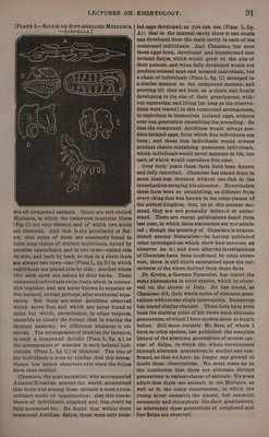
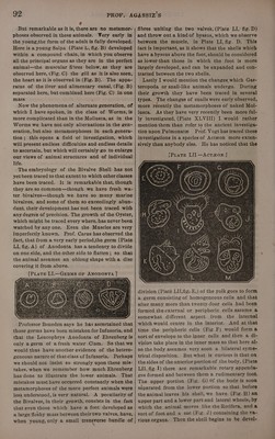
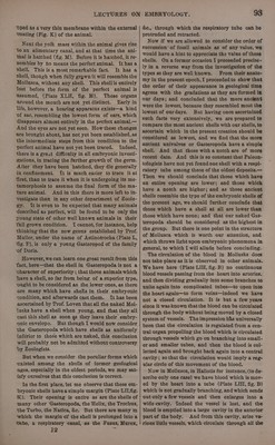
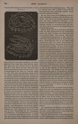
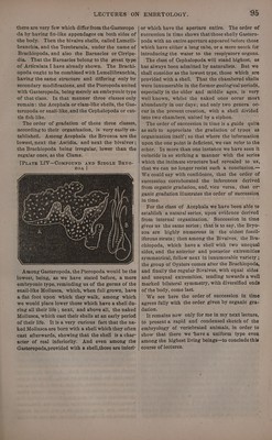
![phoses observed in these animals. Wery early in the young,the form of the adult is fully developed. Here is a young Salpa (Plate L, fig. B) developed within a compound chain, in which you observe all the principal organs as they are in the perfect animal—the muscular fibres below, as they are observed here, (Fig. C) the gill as it is also seen, the heart as it is observed in (Fig. B).. The appa- ratus of the liver and alimentary canal, (Fig. B) separated here, but combined here (Fig. C) in one mass. Now the phenomena of alternate generation, of which I have spoken, in the class of Worms, is more complicated than in the Mollusca, as in the Worms we have not only alternations in the gen- eration, but also metamorphoses in each genera- tion; this opens a field of investigation, which will present endless difficulties and endless details to ascertain, but which will certainly go to enlarge our views of animal structures and of individual life. The embryology of the Bivalve Shell has not yet been traced to that extent to which other classes have been traced. It is remarkable that, though they are so common—though we have fresh wa- ter bivalves—though we have so many marine bivalyes, and some of them so exceedingly abun- dant, their development has not been traced with any degree of precision. The growth of the Oyster, which might be traced every where, has never been watched by any one. Even the Muscles are very imperfectly known. Prof. Carus has observed the fact, that from a very early period,the germ (Plate LI, fig. A) of Anodonta has a tendency to divide on one side, and the other side to flatten; so that the animal assumes an oblong shape with a disc covering it from above. [PLate LI.—GEeRmMs OF ANODONTA |] Professor Beneden says he has ascertained that those germs have been mistaken for Infusoria, and that the Leucophrys Anodonta of Ehrenberg is only a germ of afresh water Clam. So that we would thus have another evidence of the hetero- geneous nature of thatclass of Infusoria. Perhaps we should not insist so strongly upon these mis- takes, when we remember how much Ehrenberg has done to illustrate the lower animals. That mistakes must have occurred constantly when the metamorphoses of the more perfect animals were less understood, is very natural. A peculiarity of the Bivalves, in their growth, consists in the fact that even those which have a foot developed as a large fleshy mass between their two valves, have, when young, only a small transverse bundle of fibres uniting the two valves, (Plate LI, fig. D) and throw out a kindof byssus, which we observe between the muscle, in Plate LI, fig. D. This fact is important, as it shows that the shells which have a byssus above the foot, should be considered as lower‘than those in which the foot is more largely developed, and can be expanded and con- tracted between the two shells. Lastly I would mention the changes which Gas- teropoda or snail-like animals undergo. During their growth they have been traced in several types. The changes of snails were early observed, more recently the metamorphoses of naked Mol- lusca. As they have very recently been more ful- ly investigated, (Plate XLVIII) I would rather mention them than refer to the ancient investiga- tion upon Pulmonate Prof. Vogt has traced these investigations in a species of Actzon more exten- sively than anybody else. He has noticed that the division (Plate LIL fig. E,) of the yolk goes to form a germ consisting of homogeneous cells and that after many more than twenty-four cells had been formed the external or peripheric cells assume a somewhat different aspect from the internal which would centre in the interior. And at that time the peripheric cells (Fig. F) would forma sort of envelope to the inner cells and then a di- vision take place in the inner mass so that here al- so the body assumes very soon a bilateral syme- trical disposition. But what is curious is that on the sides of the anterior portion of the body, (Plate LII, fig. 1) there are remarkable rotary appenda- ges formed and between them a rudimentary foot. The upper portion (Fig. G) Of the body is soon separated from the lower portion so that before the animal leaves his shell, we have (Fig. H) an upper part and a lower part. and lateral wheels, by which the animal moves like the Rotifera, and a sort of foot and a sac (Fig. J) containing the va- rious organs. Then the shell begins to be devel-](https://iiif.wellcomecollection.org/image/b33278982_0098.jp2/full/800%2C/0/default.jpg)