A report on the microscopic objects found in cholera evacuations / by Timothy Richards Lewis.
- Timothy Richards Lewis
- Date:
- 1870
Licence: Public Domain Mark
Credit: A report on the microscopic objects found in cholera evacuations / by Timothy Richards Lewis. Source: Wellcome Collection.
Provider: This material has been provided by Royal College of Physicians, London. The original may be consulted at Royal College of Physicians, London.
50/154 page 16
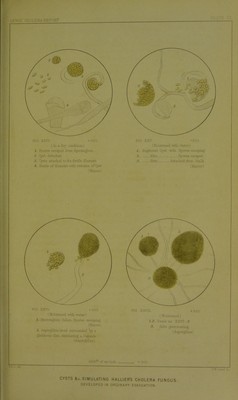
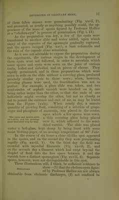
![THIS CHOLERA FUNailS OF TIIOMI?: AND Or lIALLfKR appearanco. The cells had become nearly eveiywliere atmm^ The Cholera ft^ngus of ]^SJ^'^^^,^^^^^Ongm^^^ Thome. (ingf. xxiii), coiTospondinf^ CX- actly to the figures given l)v Thome ot the cholera fungus discovered by him, to which ratlior a long name was given at the time, viz., Cylindrotccnium Cholera; Asiatkcer This condition lasted till the sixtli day, when a crop of a white mould was perceptible in the isolated preparation, and a plentiful crop of i^enicillimn and aspergillus appeared on the other cultivation (Pig. xxii). This slide having become rather dry, a few drops of distilled water were after this occasionally added. On the eighth day long delicate fila- ments were seen growing out of the ti^STr ?ei-ia^rS Y}^^^ linmus-looldng substance in first week. the apjDaratus, and on the tenth day other filaments were observed, which seemed to be tipped with various coloured heads, appa- rently of the same kind as on the other sHde, those of a bluish and yellowish-brown tint prevailing; but by the eighteenth day the long delicate filaments had grown over them, the whole surface of the preparation presenting a woolly appearance. After this no further change could be seen to take place in either cultivation, thftweX-first da^^'''' ° ^ud ou thc twcuty-fii^st day of the experiment the bell-glass was opened, and the glass plate placed on the stage of the microscope. Precisely the same species of aspergillus and penicillium were found as existed in the non-isolated cultivations, with the addition that great numbers of the filaments forming the white flocculent tuft bore at their terminations cysts or sporangia filled with distinct spores (I'ig. xxiv, 1-4), which, I think, correspond exactly to the Cysts obtained exactly like ^ysts figm^cd by Professor HalHer the cholera-cysts figured i i byHaiuer. 01 thc immature cholera-cysts, whose drawing has already been given and may be compared with this.* Aspergillus tufts were present in great numbers: nearly all of them had fallen off from A germinating aspergillus ^j^g^, fiiamcuts amoug the myccliiun; tuft simulating a cholera n t f x. cyst in the same condition, a fcw, liowcvcr, wcro pcrlcct, con- sequently easily recognised. Some * I have obtained excellent examples of this fungus (Mxicor) on the intestinal mucous membrane of the pig also, whilst subjecting strips of the intestine to contmuous (jbservatiou.](https://iiif.wellcomecollection.org/image/b24749515_0050.jp2/full/800%2C/0/default.jpg)