Atlas and epitome of ophthalmoscopy and ophthalmoscopic diagnosis / by O. Haab.
- Haab, O. (Otto), 1850-1931.
- Date:
- 1910
Licence: In copyright
Credit: Atlas and epitome of ophthalmoscopy and ophthalmoscopic diagnosis / by O. Haab. Source: Wellcome Collection.
44/376 (page 40)
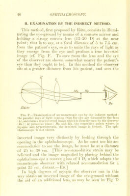
![II. EXAM1NATION BY THE INDIRECT METHOD. This method, first proposed by Rüte, consists in illumi- nating the eye-ground by means of a concave mirror and holding a strong convex lens (13-20 D) at the near point; that is to say, at a focal distance of 5 to 7.5 cm. from the patient’s eye, so as to nnite the rays of light as they emerge from the eye and produce a trne inverted image (cf. Fig. F. To save room the lens and the eye of the observer are shown somewhat nearer the patient’s eye than they ought to be). In this method the observer sits at a greater distance from bis patient, and sees the Fig. F.—Examination of an emmetropic eye by the indirect method : the parallel rays of light Corning from the eye are focussed hy the lens (L), and form a true inverted image which is directly seen by the observer (Be.); II, principal plane; My. and Ily., the respective poiuts where, in myopia and hypermetropia, the inverted image is formed. The oph- thalmoscope is not shown. inverted image very distinctly by looking through the opening in the ophthalmoscope. As he must use his ac- commodation to see the image, he must be at a distance of 25 to 30 cm. [This accommodative strain may be relieved and the image magnified by placing behind the ophthalmoscope a convex glass of 4 D, which adapts the emmetropic observer with relaxed accommodation for a point 25 cm. distant.—Ed.] In high degrees of myopia the observer can in this way obtain an inverted image of the eye-ground without the aid of an additional lens, as may be seen in Fig. D](https://iiif.wellcomecollection.org/image/b28128655_0044.jp2/full/800%2C/0/default.jpg)