Licence: Public Domain Mark
Credit: Embryology : from Quain's anatomy, ninth edition. Source: Wellcome Collection.
Provider: This material has been provided by The University of Glasgow Library. The original may be consulted at The University of Glasgow Library.
171/196
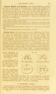
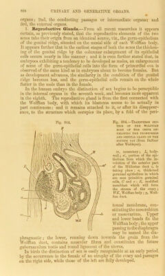
![Urinary Bladder and Urachus.—The urinary bladder, as already stated in an earlier part of this description, is formed by a dilatation of the stalk of the allantois. In the human foetus this appears as a spindle-shaped cavity at the end of the second month, the narrower part below being prolonged as the first part of the urethra into the cloaca, while the upper narrowing part extends as the urachus into the umbilical cord, and at an early period in animals is prolonged into the cavity of the allantois. The ureters terminate in the dorsal wall of that part of the urogenital sinus which is dilated into the urinary bladder. The urachus is usually a solid cord at a short distance from the bladder, but, according to Luschka, it not unfrequently remains hollow for some length within the umbilical cord. The spindle shape of the bladder is retained for a long time in tho human foetus. For the development of the Suprarenal bodies, see p. S41. Genital Cord.—In both sexes, as was first, tally shown by Tiersch (No. 287) and Lenckart (No. 287*) in 1802, the two Wolttian ducts are united by surrounding substance into one cord behind what becomes the base of the urinary bladder ; but retaining internally their separate passages until they reach the sinus urogeuitalis. With this cord the Mullerian duets are incorporated posteriorly, so that at one time there are four passages through the whole of the genital coni. The MUllerian ducts next coalesce into one at wane little distance from their lower ends, and this fusion progressing upwards and downwards for a considerable space, a single median cavity is produced which lies between the still separate canals of the Wolffian ducts. A large accumulation of tissue in its walls gives to the genital cord great thickness as compared Fig. 803.—Transverse SEC- TIONS OK T1IK GKXITAL COR!) IS A FEMALE CALK KMHHYO. Magnified 14 diameters (from KiiUiker). 1, near the upper end ; 2 and 3, near the middle ; 4, at the lower end ; a, anterior, ]>, posterior aspect; m, Mul- lerian ducts united or sei«t- rnte; w, Wolffian ducts. with the neighbouring parts of the ducts where they emerge from its enclosure. The lower Fig. 803. part of the united Mullerian ducts thus comes afterwards to form the foundation of the vagina and lower part of the uterus in the female, and the corresponding prostatic vesicle with its occasional vestigial accompaniments in the male, or the uterus masculinus. IX. GENERATIVE ORGANS. Tn the history of the further development of the generative organs it wdl be convenient to consider them in the two sexes in succession under t io three heads of, 1st, the reproductive glauds, or strictly internal vol. nt * 3 M](https://iiif.wellcomecollection.org/image/b24931512_0175.jp2/full/800%2C/0/default.jpg)