A report of surgical cases treated in the Army of the United States from 1865 to 1871 / War Department, Surgeon General's Office.
- Date:
- 1871
Licence: Public Domain Mark
Credit: A report of surgical cases treated in the Army of the United States from 1865 to 1871 / War Department, Surgeon General's Office. Source: Wellcome Collection.
Provider: This material has been provided by the Royal College of Physicians of Edinburgh. The original may be consulted at the Royal College of Physicians of Edinburgh.
282/318 page 266
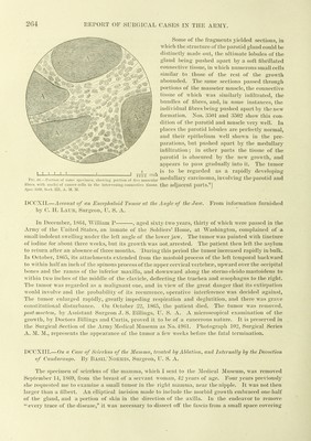
![r 266 EEPOET OF SURGICAL CASES IN THE AR]\IY. Army Medical Museum, Wmlu))f/tnn, J). C, July 21, 1871. Sir : In accordance with your request, I have made a niiscroscopical exauiination of the several morbid growths in the case of Isabella G , with the general conclusion that the case Avas undoubtedly one of scirrhus carcinoma. The specimens preserved at the Museum are as follows: No. 5598, Surgical Section. The tumor first removed by Surgeon Norris is an oval mass, about five inches long by three broad, with a portion of skin attached externally. The greater portion of the piece is occupied by a firm irregular scirrhus growth, which, after being several months in alcohol, still yielded, on scraping, a creamy juice containing cells and nuclei; on tlie edges of the mass is some normal adipose tissue. No. 5921, Surgical Section. The tumor removed by Surgeon Norris at the second operation is a smaller mass, containing a similar but somewhat softer growth. In the skin, adherent exter- nally, a portion of the cicatrix of the first operation can be recognized, and from this an irregular soft nodule, the size of a walnut, protrudes. No. 10S3, ]Medical Section, is a portion of the stomach and the spleen of the same patient. In the greater curvature of the stomach, and not far from the cardiac orifice, is a carcinomatous thickening of the coats of the organ, about half an inch in thickness, occupying an area about four inches in diameter. Externally the thick- ened patch was closely connected with the hilus of the very small spleen by a soft carcinomatous tumor, about the size of a hen's egg. Sections of the first and second tumors re- moved from the breast, of the stomach, and the * spleen, were ])repared in the Microscopical Sec- tion of the Museum, by Acting Assistant Sur- geon E. M. Schaeffer, and having been stained with carmine and mounted in Canada balsam, now form a part of the microscopical collection. Nos. 3513 to 3519, Microscopical Section, are from the tumor first removed from the breast, and show it to consist of an areolar stroma of soft connective tissue, the inter- spaces of which are stuffed with masses of loosely adherent, nucleated cells; the nuclei of these cells are oval, and measure from three to sixten-thousandths of an inch in long diameter; they contain generally one or two shining nucleoli. The cells themselves are of an irregular form, and various sizes, the smaller ones predominating. The connective tissue stroma contained an abundance of small nucleated, connective tissue coi-puscles, resembling those found in most new formations of connective tissue. Nos. 3G73 to 3G76, Microscopical Section, are from the second breast tumor, and are very similar to those from the first; in many places, however, the cell masses are more voluminous and the con- nective tissue stroma is less prominent. Nos. 36G3 to 3672, Microscopical Section, are sections cut perpendicularly to the surface, through the indurated portion of the stomach. In most places the mucous membrane and the tubular glands are in a condition not far from normal; but the submucous connective tissue, the muscular and iieritoneal coats, are transformed into a carcinomatous mass, consisting of grouj^s of cells with large nuclei imbedded in a connective tissue stroma, the general character of the neoplasm closely approximating those of the breast tumors. Nos. 3656 to 3662, Microscopical Section, are sections of tumor between the spleen and the stomach; they present the same general characteristics, but this tumor was much softer than the other growths, and in the section the stroma appears less pronounced, and the cell masses are more voluminous. In this case, as in others of multiple carcinoma, examined in the Microscopical Section Fig. 67.—Section of scinlius, magnified 400 diameters. »S^jjcc. 3513,](https://iiif.wellcomecollection.org/image/b21970695_0282.jp2/full/800%2C/0/default.jpg)