Genetic recombination : understanding the mechanisms / Harold L.K. Whitehouse.
- Harold Leslie Keer Whitehouse
- Date:
- [1982]
Licence: Attribution-NonCommercial-NoDerivatives 4.0 International (CC BY-NC-ND 4.0)
Credit: Genetic recombination : understanding the mechanisms / Harold L.K. Whitehouse. Source: Wellcome Collection.
59/448 page 45
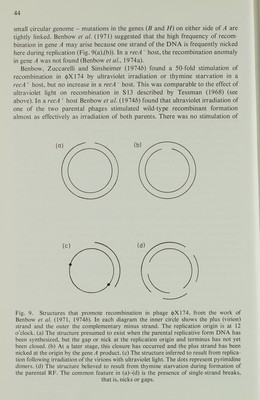
![recombination by ultraviolet irradiation in the high recombination region of gene A. This suggests that the ultraviolet irradiation effect and the gen eA effect involve the triggering of the same mechanism, the ultraviolet light in consequence being unable to increase recombination further. Benbow et al. (\914b) examined by various techniques the structure of the RF produced on infecting E. coli with ultraviolet-irradiated <j>X174. The normal RF is of two kinds: covalently closed supercoiled duplex circles called RFI, and relaxed duplex circles called RFII; these have one or more nicks or gaps. On a neutral sucrose velocity sedimentation gradient, the RFI and RFII sediment at different positions. When replication of the parental RF was blocked by treatment with chloramphenicol or in other ways, 65% of the DNA sedimented as RFI and the remainder as RFII. With increasing ultraviolet radiation dose to the parent phage the proportion of the parental RF sedimenting at the RFII position increased. In addition, many ultraviolet-damaged molecules sedimented anomalously. Similar results were obtained whether the host was recA + or recA except that with recA + 20% of the DNA sedimented at a position indicative of multiple-length molecules. In an alkaline sucrose velocity gradient, molecules other than RFI are denatured. With ultraviolet-irradiated phage, apart from some RFI molecules, the viral (plus) strands (which had been labelled with [ 14 C]thymidine) sedimented primarily as full length circles, while the complementary (minus) strands (which had been labelled with [ 3 H]thymidine) sedimented as linear molecules of various sizes but shorter than full length. These results, both for the neutral and the alkaline gradients, confirm those of Francke and Ray (1971) for a recA + host. Sedimenta tion of the doubly labelled RF on a neutral gradient showed less 3 H than 14 C in the RFII when the parent phage had been treated with ultraviolet light. This result indicates that the minus strands were incomplete. All the sedimentation data fit the hypothesis that, following ultraviolet irradiation of the phage, the irradiated plus strand remains as an intact circle, but synthesis of the complementary minus strand proceeds from the replication origin but stops at the first pyrimidine dimer or other ultraviolet-induced lesion which it encounters in the plus strand template (Fig. 9(c)). Electron microscopy of parental RF molecules spread in an aqueous film of cytochrome c revealed one duplex and one simplex region within each circular molecule, following ultraviolet irradiation of the <j)X174 particles. The length of the duplex region was inversely related to the ultraviolet dose. These findings are in keeping with the conclusions from the sedimentation data. The experiments on the effect of thymine starvation were carried out by infect ing a thymine-requiring {thy') host strain with the phage and growing the cells for 20 min in a medium lacking thymine. The data for the parental RF from a neutral sucrose gradient, using thymine-starved cells, were found to be similar to those for ultraviolet-irradiated phage, but the alkaline gradient data showed 3 H- and Re labelled DNA principally as linear molecules of up to full length. Under the electron microscope the molecules were seen to be predominantly circular duplex RFII (or at least of RFII appearance) of normal length, but 10-20% were linear, also of full length. Simplex regions of more than 100 nucleotides would have been](https://iiif.wellcomecollection.org/image/b18020768_0060.JP2/full/800%2C/0/default.jpg)
 No text description is available for this image
 No text description is available for this image
 No text description is available for this image