On deformity of the lower jaw in the cachalot (Physeter macrocephalus, Linn.) / by James Murie.
- Murie, James.
- Date:
- [1865]
Licence: Public Domain Mark
Credit: On deformity of the lower jaw in the cachalot (Physeter macrocephalus, Linn.) / by James Murie. Source: Wellcome Collection.
Provider: This material has been provided by The Royal College of Surgeons of England. The original may be consulted at The Royal College of Surgeons of England.
6/12 page 392
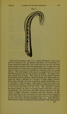
![the inner surface of the bone, which ought to have been in approxima- tion with the left half, comes to be upon the upper surface (fig. 1 b), while the sockets are thrown round, partly looking opposite to those in the hinder part of the jaw, and partly on the under surface. The bone in this instance is increased in density, and several of the sockets for the teeth have spongy bone thrown out upon their sur- faces, showing that the parts have suffered at one time or other from chronic inflammation. The bone otherwise is healthy, or such as if it had not suifered from rachitis or other softening causes. The third example is one which gives a better idea of this anoma- lous condition (see fig. 2). It is a specimen contained in the Osteo- logical Collection at the British Museum, which I was enabled to examine carefully through the kindness of Dr. Gray and Mr. Gerrard. There is no history attached to it. The two halves of the jaw are complete, but separated from each other. Their size shows the animal to have been young, although of considerable dimensions. The length of the two, placed in juxta- position, in a straight median line from opposite the posterior ends of the rami to the anterior surface of the bend, is about 65 inches, while the measurement following the curve of the right half to the tip of the jaw is 92 inches. Their anterior fourth has a curve to- wards the left side, in shape not unlike a shepherd's crook ; and they have besides a twist upon themselves. Each lateral half of this inferior maxillary bone presents characters sufficient to make it worthy of a separate description. The left has twenty-two sockets in its alveolar process. Following these as the most simple guide to the nature of the twist, we find the posterior six alveoli to be nearly on the upper surface, or in natural position; the next seven in advance (which occupy the hinder end of the sym- physis) by degrees change from the upright position, so that the foremost one at the middle of the crook comes to be on the outer side of the jaw, and points directly backwards to the condyles. The four alveoli anterior to these last return gradually from the outer side to the upper surface, and the remaining five in front continue as it were in their normal position, that is, directed upwards. The bone of the jaw, besides the double angular bend, has in the mean time rolled itself outwards along with the sockets, so that the internal edge of the symphysis at the sharp bulging curve (fig. 2 a) is upon the upper surface; then as the bone bends backwards and outwards the symphysis returns or rolls itself inwards (fig. 2 e), so that at tlie anterior end it is first above and then comes to be almost on the inward and under surface. The symphysis has therefore a double bend and a double twist. The pathological condition of the left half of the jaw is as fol- lows :—The alveoli, as far forwards as the wide bend, are partially filled with spongy exostosed bone.; the rest of the anterior alveoli have likewise traces of spongy bone in them, but their cavities seem rather widened than otherwise. The bone of the symphysis at the outward bend is very much augmented in breadth, thickness, and density (fig. 2 a); and internally it fits into a large hollow in the [4]](https://iiif.wellcomecollection.org/image/b2228672x_0006.jp2/full/800%2C/0/default.jpg)