Volume 1
A text-book of human physiology : including histology and microscopical anatomy with special reference to the requirements of practical medicine / by L. Landois ; translated from the seventh German edition with additions by William Stirling.
- Landois, L. (Leonard), 1837-1902. Lehrbuch der Physiologie des Menschen. English
- Date:
- 1891
Licence: Public Domain Mark
Credit: A text-book of human physiology : including histology and microscopical anatomy with special reference to the requirements of practical medicine / by L. Landois ; translated from the seventh German edition with additions by William Stirling. Source: Wellcome Collection.
129/602 page 89
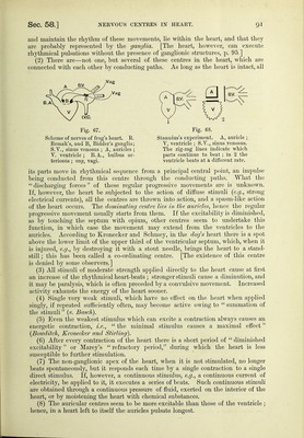
![done through a certain thickness of lung tissue, and hence one must strike the pleximeter forcibly. It extends vertically from the third rib and ends at the sixth, but owing to the cardiac merging in the hepatic dulness, this lower limit cannot be accurately ascertained ; while transversehj at the fourth rib it extends from just within the nipple line to slightly beyond the right of the sternum. By these means we may detect increase in the size of the heart or altera- tions in the relation of the lungs to the heart, fluid in pericardium, &c. Thus it is of great importance to the clinician, enabling him to determine the size and position of the heart.] [Auscultation.—This is one of the most valuable methods, for by it we can detect variations and modifications in the healthy sounds of the heart, the rhythm and frequency of the heart- beat, the existence of abnormal sounds, and their exact relation to the normal sounds, also their characters and relation to the cardiac cycle, and the direction in which these sounds are propagated (§ 54).] 57. INNERVATION OF THE HE ART. —[Intra- and Extra-Cardiac Nervous Mechanism.—Wlien tlie lieart is removed from tlie body, or wlien all ttie nerves whicli pass to it are divided, it still beats for some time, so that its movements must depend upon some mechanism situated within itself. The ordinary rhythmical movements of the heart are undoubtedly associated with the presence of nerve ganglia, which exist in the substance of the heart—the intra-cardiac ganglia. But the movements of the heart are influenced by nervous impulses which reach it from without, so that there falls to be studied an intra-cardiac and an extra-cardiac nervous mechanism.] The cardiac plexus is composed of the following nerves :—(1) The cardiac branches of the vagus, the branch of the same name from the external branch of the superior laryngeal, a branch from the inferior laryngeal, and sometimes branches from the pulmonary plexus of the vagus (more numerous on the right side); (2) the superior, middle, inferior, and lowest cardiac branches of the three cervical ganglia and the first thoracic ganglia of the sympathetic ; (3) the inconstant twig of the descending branch of the hypoglossal nerve, which, according to Luschka, arises from the upper cervical ganglion. From the plexus there proceed—the deep and the superficial nerves (the latter usually at the division of the pulmonary artery under the arch of the aorta, and containing the ganglion of Wrisberg) (§ 370). The following nerves may be separately traced from the plexus :— (a) The plexus coronarius dexter and sinister, which contains the vaso-motor nerves for the coronary vessels (physiological proof still wanting) as well as the nerves (sensory ?) proceeding from them (to the pericardium ?). {h) Intra-cardiac nerves and ganglia.—The nerves lying in the grooves of the heart and in its substance contain numerous ganglia {Remak), and are regarded as the automatic motor centres of the heart. A nervous ring containing numerous ganglia corresponds to the margin of the septum atriorum ; there is another in the auriculo-ventricular groove.' Where the two meet, they exchange fibres. The ganglia usually lie near the pericardium. In mammals, the two largest ganglia lie near the orifice of the superior vena cava—in birds, the largest ganglion (containing thousands of ganglionic cells) lies posteriorly where the longitudinal and transverse sulci cross each other. Fine branches, also provided with small ganglia, proceed from these ganglia, and penetrate the muscular walls of the auricles and ventricles. [Frog's Heart.—The frog's heart consists of the sinus venosus, into which open the single inferior and the two superior venae cavse (fig. 64). There are two auricles ; the right one com- municates with the sinus venosus, and opens into the single ventricle ; the left auricle also opens into the single ventricle (fig. 63, v), and in the latter are mixed the venous blood returned by the right auricle and the arterial blood from the left auricle. The aorta with its hulhus arteriosus conducts the blood from the ventricle. The various orifices are guarded by projec- tions of tissue, which act like valves. The two auricles are completely separated by a septum. This septum ends posteriorly in a free concave margin, so as to divide the auriculo-ventricular orifice into a right and left orifice. Each orifice is guarded by two thick fleshy valves, which close it.] [Nerves.—The two cardiac branches of the vagi—the nervi cardiaci—proceed to the posterior surface of the sinus venosus, and where the latter joins the auricle they interlace, and are mixed](https://iiif.wellcomecollection.org/image/b20417688_001_0129.jp2/full/800%2C/0/default.jpg)


 No text description is available for this image
 No text description is available for this image
 No text description is available for this image