Volume 1
A text-book of human physiology : including histology and microscopical anatomy with special reference to the requirements of practical medicine / by L. Landois ; translated from the seventh German edition with additions by William Stirling.
- Landois, L. (Leonard), 1837-1902. Lehrbuch der Physiologie des Menschen. English
- Date:
- 1891
Licence: Public Domain Mark
Credit: A text-book of human physiology : including histology and microscopical anatomy with special reference to the requirements of practical medicine / by L. Landois ; translated from the seventh German edition with additions by William Stirling. Source: Wellcome Collection.
583/602 page 543
![Historical.—Aristotle directed attention to the relatively large size of the human bladder— he named the ureters. Massa (1552) found lymphatics in the kidney. Eustachius (f 1580) ligatured the ureters and found the bladder empty. Cusanus (1565) investigated the colour and weight of the urine. Rousset (1581) described the muscular nature of the walls of the bladder. Yesling described the trigone (1753). The first important chemical investigations on the urine date from the time of van Helmont (1644). He isolated the solids of the urine, and found among them common salt; he ascertained the higher specific gravity of fever-urine, and ascribed the origin of urinary calculi to the solids of the urine. Scheele (1766) discovered uric acid and calcium phosphate ; Arandand Kunckel, phosphorus ; Rouelle (1773), urea ; and it got its name from Fourcroy and Vauquelin (1799). Berzelius found lactic acid ; Seguin, albumin in pathological urine ; Liebig, hippimc acid ; Heintz and v. Pettenkofer, kreatin andkreatinin; Wollaston (1810), cystin. Marcet found xanthin ; and Lindbergson, magnesic carbonate. 283. STRUCTURE OF THE SKII^, HAIRS, AND NAIL.—The skin (3-3 to 2*7 mm. thick; specific gravity, 1057) consists of— [1. The epidermis; 2. The chorium, or cutis vera, with the papillse (fig. 356).] The epidermis (0-08 to 0-12 mm. thick) consists of many layers of stratified Functions of the Skin. ^'H^ , Epidermis. % , v'f 1^^^ Eleidin -^ ' ^/ layer. 4'&' Stratum ''W' Malpigliii. Papilla. -2 ,f- Fat-cells. Sweat-duct. Fig. 356. Vertical section of the human skin. epithelial cells united to each other by cement substance (figs. 356, 357, 358). The superficial layers—stratum corneum—consist of several or many layers of](https://iiif.wellcomecollection.org/image/b20417688_001_0583.jp2/full/800%2C/0/default.jpg)

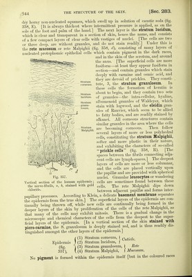


 No text description is available for this image
 No text description is available for this image
 No text description is available for this image