Volume 1
A text-book of human physiology : including histology and microscopical anatomy with special reference to the requirements of practical medicine / by L. Landois ; translated from the seventh German edition with additions by William Stirling.
- Landois, L. (Leonard), 1837-1902. Lehrbuch der Physiologie des Menschen. English
- Date:
- 1891
Licence: Public Domain Mark
Credit: A text-book of human physiology : including histology and microscopical anatomy with special reference to the requirements of practical medicine / by L. Landois ; translated from the seventh German edition with additions by William Stirling. Source: Wellcome Collection.
587/602 page 547
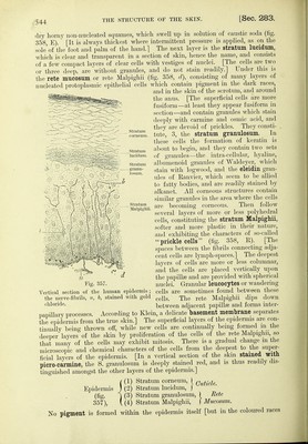
![same time, the matrix, from wliicli growth of the nail takes place. The liuiule is present in an isolated nail, and is due to diminished transparency of the posterior part of the nail, owing to the special thickness and uniform distribution of the cells of the rete Malpighii {Toldt). Growth of the Nail.-Accordin^ to Unna, the matrix extends to the front part of the lunula The nail grows continually from behind forwards, and is formed by layers secreted or formed by the matrix. These layers run parallel to the surface of the matrix. They run obliquely fiom above and behind, downwards and forwards, through the thickness of the siibstance t the nail The nail is of the same thickness from the anterior margin of the lunule forwards to its free margin. Thus the nail does not grow in thickness in this region. In the course ot a year the fingers produce about 2 grms. of nail substance, and relatively more m summer than m Dlvelopment.-l. From the second to the eighth month of fffital life, the position of the nail is indicated by a partial but marked horny condition of the epidermis on the back ot the first phalanx, the eponychium. The remainder of this substance is represented during life by the normally formed epidermal layer, which separates the future nail from the surface ot the furrow 2. The future nail is formed under the eponychium, with its first nail-cells still m tront of the nail-groove ; then the nail grows and pushes forward towards the groove. At the seventh month, the nail (itself covered by the eponychium) covers the whole extent of the nail-bed. 3. When, at a later period, the eponychium splits off, the nail is uncovered. After birtti the ridges are formed on the bed of the nail, while simultaneously the matrix passes backwards to the'^most posterior part of the groove (f/?ma). Absence of Hairs.—The whole of the skin, with the exception of the palmar surtace ot the hand, sole of the foot, dorsal surface of the third phalanx of the fingers and toes, outer surface of the eyelids, glans penis, inner surface of the prepuce, and part of the labia, is covered with hairs, which may be strong or fine (lanugo). A Hair (specific gravity 1-26) is fixed by its lower extremity (root) in a depression of the skin or a hair-follicle (fig. 358, I, p) which passes obliquely through the thickness of the skin, sometimes as far as the subcutaneous tissue. The structure of a hair-follicle is the following :—1. The outer fibrous layer (figs. 358, 1, 357), composed of interwoven bundles of connective-tissue, arranged for the most part longitudinally, and provided with numerous blood-vessels and nerves. [It is just the connective-tissue of the sur- rounding chorium.] 2. The inner fibrous layer (figs. 358, 2, 361) consists of a layer of fusiform cells (? smooth mus- cular fibres) arranged circu- larly. [It does not extend throughout the whole length of the follicle.] 3. Inside this layer is a transparent, hyahne, glass-like base- ment membrane (fig. 358, 3, 361), which ends at the neck of the hair-foUicle; while above it is continued as the basement membrane which exists between the epidermis and chorium (?). In addition to these coverings, a hair-follicle has epithelial coverings which must be regarded in relation to the layers of the epidermis. Immediately within the glass-like membrane is the outer root-sheath (figs. 358, 4, 361, 362), which consists of so many layers of epitheHal cells that it forms a conspicuous covering. It is, in fact, a direct continuation of the stratum Malpighii, and consists of many layers of soft cells, the cells of the outer layer a, nail-substance; Fig. 360. Transverse section of one-half of a nail. b, more open layer of cells of the nail-bed ; c, stratum Malpighii of the nail-bed ; d, transversely divided ridges ; nail-groove ; /, horny layer of e projecting over the g, papilltB of the skin on the back of the finger. nail](https://iiif.wellcomecollection.org/image/b20417688_001_0587.jp2/full/800%2C/0/default.jpg)


 No text description is available for this image
 No text description is available for this image
 No text description is available for this image