The law of cardiac muscle with special reference to conduction in the mammalian heart / Thomas Lewis.
- Thomas Lewis
- Date:
- [1921]
Licence: Public Domain Mark
Credit: The law of cardiac muscle with special reference to conduction in the mammalian heart / Thomas Lewis. Source: Wellcome Collection.
5/18 page 341
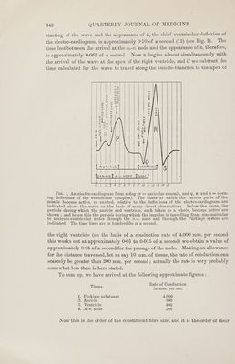
![We will now proceed to examine and compare the power of these four tissues to conduct. Rates of Conduction. Many observations have been undertaken upon rates of conduction in the heart muscle; many of the methods used have been faulty or subject to very appreciable error. That has been so because the paths of conduction have been recognized only recently, and no observations upon rate of conduction can be of value which do not take into account the path followed by the wave. The statements which follow apply to the dog’s heart. In the awricle. In the mammalian auricle the natural rate of conduction is estimated with precision by first ascertaining the exact starting-point of the wave and its lines of propagation, and by subsequently measuring the times of its arrival at points on these lines. That can be, and has been, done both for the excitation wave and the contraction wave which follows it (8). The rate is the same for both processes, and approaches the average figure of 800 mm. per second. The rate of propagation may also be ascertained by stimulating the auricle rhythmically in line with two points at which the times of arrival of the propagated waves are signalled. Values similar to those obtained for the natural wave are found (9). In the ventricle. The mammalian ventricle offers peculiar difficulties, because the rates of conduction in the ventricular muscle and in the Purkinje substance, which lines its endocardia] surface, are by no means the same. The method of chief value is to propagate waves artificially from the pericardial surface of the ventricle and in line with two points at which the arrival of the resultant waves is timed. Two precautions are essential to accuracy. The muscular wall must be of considerable thickness; for this reason the left ventricle is chosen; and the point of stimulation and points signalling the waves must be close together. Otherwise the rate of conduction obtained is not that of ventricular muscle, but is a rate comprised of that of ventricular muscle and Purkinje substance. The rates of both are measurable with considerable accuracy by adopting suitable methods; that of ventricular muscle averages 400 mm. per second, that of straight Purkinje fibres is approximately 4,000 mm. per second (10). In the a-v. node. A direct measure of conduction in the node is not possible ; that conduction is slow is suspected from the delay between the contractions of auricle and ventricle, and from analogy with the fibres of the a.-v. ring of the frog. An estimate of actual conduction rate in the node may be obtained by considering certain transmission intervals. In the naturally beating heart of medium-sized dogs the transmission interval from the s.-a. node to the septal muscle overlying the a.-v. node measures approximately 0-03 to 0-04.0f a second. By a different method the interval to the a.-v. node itself is estimated at similar figures. We may take the mean 0-035 of a second as an estimate subject to no great error (11). The interval between the](https://iiif.wellcomecollection.org/image/b33449168_0005.jp2/full/800%2C/0/default.jpg)