The law of cardiac muscle with special reference to conduction in the mammalian heart / Thomas Lewis.
- Thomas Lewis
- Date:
- [1921]
Licence: Public Domain Mark
Credit: The law of cardiac muscle with special reference to conduction in the mammalian heart / Thomas Lewis. Source: Wellcome Collection.
6/18 page 342
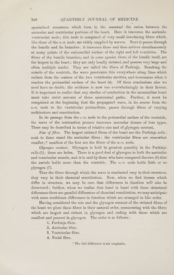
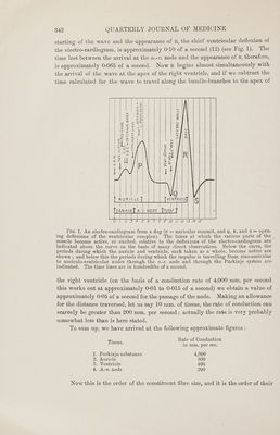
![~ starting of the wave and the appearance of R, the chief ventricular deflexion of the electro-cardiogram, is approximately 0-10 of a second (12) (see Fig. 1). The time lost between the arrival at the a.-v. node and the appearance of R, therefore, is approximately 0-065 of a second. Now R begins almost simultaneously with the arrival of the wave at the apex of the right ventricle, and if we subtract the time calculated for the wave to travel along the bundle-branches to the apex of AURICLE Rey al { | 1 Al | | $v 44 | Be | a lena es : | a 2= = We ais = < ee = 22 <o v Bs eel c lg eee, ona ee Gd reas] | ae aay | x e= = 5 oe, ] k el ene uy q ie (i (a fied | Go igs 2 | Bs OS Posy : Te) ware fi a 7 29 | Sel | IRI fete va <7 Ces <= | zk Ms ww | | > t a 4 \ 4 : | | tS AN-AVN. ay NODE CRN Iu PS SOR TUE RS OLS ' ’ _ Fie. 1. An electro-cardiogram from a dog (P = auricular summit, and Q, R, and S = open- ing deflexions of the ventricular complex). The times at which the various parts of the ~ muscle become active, or excited, relative to the deflexions of the electro-cardiogram are indicated above the curve on the basis of many direct observations. Below the curve, the periods during which the auricle and ventricle, each taken as a whole, become active are shown ; and below this the periods during which the impulse is travelling from sino-auricular to auriculo-ventricular nodes through the a.-v. node and through the Purkinje system are indicated. The time lines are in hundredths of a second. the right ventricle (on the basis of a conduction rate of 4,000 mm. per second this works out at approximately 0-01 to 0-015 of a second) we obtain a value of approximately 0-05 of a second for the passage of the node. Making an allowance for the distance traversed, let us say 10 mm. of tissue, the rate of conduction can scarcely be greater than 200 mm. per second; actually the rate is very probably somewhat less than is here stated. To sum up, we have arrived at the following approximate figures : Rate of Conduction Tissue. in mm. per sec. 1. Purkinje substance 4,000 2. Auricle 800 3. Ventricle 400 4, A.-v. node 200 Now this is the order of the constituent fibre size, and it is the order of their](https://iiif.wellcomecollection.org/image/b33449168_0006.jp2/full/800%2C/0/default.jpg)