Principles of human physiology / by William B. Carpenter ; edited by Henry Power.
- Date:
- 1876
Licence: Public Domain Mark
Credit: Principles of human physiology / by William B. Carpenter ; edited by Henry Power. Source: Wellcome Collection.
Provider: This material has been provided by the Royal College of Physicians of Edinburgh. The original may be consulted at the Royal College of Physicians of Edinburgh.
22/1238
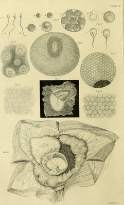
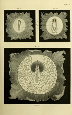
![XVI EXPLANATION OF PLATES. FIG. 9. PortioD of the mucous layer of the germinal membrane, highly magnified ; showing that it is made-up of cells, whose borders are more distinct and more closely applied to each other than those of the serous layer, and whose contents are more transparent (§ 776). [The six preceding figures are after Bischoff (“ Entwickelungsgeschichte der Siiugethiere,” &c. (1842),— “ des Kaninchen-eies”—(1842)—“desHunde- eies” (1845).] 10. Gravid Uterus of a Woman who had committed suicide in the seventh week of pregnancy, laid open :—a, os uteri internum ; 6, cavity of the cervix ; c, c, c, c, the four flaps of the body of the uterus turned back ; d, d, d, inner surface of uterine decidua ; e, e, decidua reflexa; /, /, external villous surface of the chorion; g, internal surface of the chorion; h, amnion ; i, umbilical vesicle; Tc, umbilical cord; l, embryo ; m, space between chorion and amnion (§ 752 et seq.). [After Wagner (“leones Physio- logic®”).] PLATE II. 11. Uterine Ovum of Rabbit, showing the Area Pellucida, with the primitive trace (§ 776). 12. More advanced Ovum, showing the incipient formation of the Vertebral column, and the dilatation of the primitive groove at its anterior extremity (§ 776). 13. More advanced Embryo, seen on its ventral side, and showing the first develop- ment of the Circulating apparatus. Around the Vascular Area is shown the terminal sinus a, a, a. The blood returns from this by two superior branches, b, b, and two inferior, c, c, of the omphalo-mesaraic veins, to the heart, d ; which is, at this period, a tube curved on itself, and pre- senting the first indication of a division into cavities. The two aortic trunks appear, in the abdominal region, as the inferior vertebral arteries, e, e ; from which are given off the omphalo-mesaraic arteries /, /, which form a network that distributes the blood over the vascular area. In the cephalic region are seen the anterior cerebral vesicles, with the two ocular vesicles, g (§ 777). [The three preceding figures are from the woi'ks of Bischoff previously cited.] PLATE III. (To face page 28.) Comparative View of the Skeleton of Man, and that of the Orang Outan. [After Owen (“Zoological Transactions,” vol. i.).]](https://iiif.wellcomecollection.org/image/b21965237_0022.jp2/full/800%2C/0/default.jpg)