A manual for the student of anatomy : containing rules for displaying the structure of the body, so as to exhibit the elementary views of anatomy and their application to pathology and surgery / by John Shaw.
- John Shaw
- Date:
- 1821
Licence: Public Domain Mark
Credit: A manual for the student of anatomy : containing rules for displaying the structure of the body, so as to exhibit the elementary views of anatomy and their application to pathology and surgery / by John Shaw. Source: Wellcome Collection.
Provider: This material has been provided by King’s College London. The original may be consulted at King’s College London.
139/372 page 117
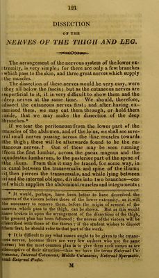
![■ for it almost immediately passes through the inlerosseu* • ligament; but by raising the fascia which covers the deep j layer of muscles, we shall see the posterior tibiae, i through almost its whole course. This artery generally ■ .gives off the peroneal, or fibular artery, about half : an inch, or an inch, below the edge of the popliteus ! muscle. But the fibular is very irregular; indeed it is ! described, by many, as rising more frequently from the I anterior, than the posterior tibia]. While the posterior j tibial is passing the insertion of the popliteal muscle, it I .gives off a branch, which, passing into the bone, is called 1 the JV'utritia Tibiae. The artery may then be traced, under j the fascia, to below the inner ankle, without our seeing j any branch of importance; but here it sends some i branches to the heel, which are called Calcanea?, and then i divides into the plantaris externa and plantaris in- i terna,—which are to be carefully traced between the muscles in the sole of the foot: in doing this, we shall be obliged to cut many of the muscles. The plantar arteries will be seen to form inosculations with those branches of the anterior tibial, which perforate the spaces between the metatarsal bones. We should now return to the dissection of the branches of the fibular artery. This vessel is not only very ir- regular in its origin, but also in its size; for it always de- pends upon the magnitude of the anterior and posterior tibial arteries. In its course towards the ankle, it gives off small branches to the muscles rising from the fibula,— one to the bone itself; and when about four inches from the ankle, it will be found to divide into two branches, which are called Anterior Fibular and Posterior Fibular. The anterior inosculates with the branches from the Tar- seal of the anterior tibial, while the posterior inosculates with the Calcanece of the posterior tibial. We may now make the dissection of the anterior tibial. To find it, we should first expose the muscles on the fore part. In doing this, we shall see the recurrens passing back upon the knee; then, by dissecting between the tibialis anticus and extensor communis digitorum, We •shall discover the main artery, lying close upon the inter- osseous ligament. It may then be easily traced to the .great toe, giving off branches in its course, the names of which are descriptive of the parts which they supply. The manner of dissecting the arteries, which has just ibeen described, should be nearly followed in making a (preparation; but the dissection should be prosecuted in a very different manner, in studying the surgical anatomy :](https://iiif.wellcomecollection.org/image/b21305158_0139.jp2/full/800%2C/0/default.jpg)
 No text description is available for this image
 No text description is available for this image
 No text description is available for this image