Atlas of urinary sediments : with special reference to their clinical significance / by Hermann Rieder ; translated by Frederick Craven Moore ; edited and annotated by A. Sheridan Delépine.
- Hermann Rieder
- Date:
- 1899
Licence: Public Domain Mark
Credit: Atlas of urinary sediments : with special reference to their clinical significance / by Hermann Rieder ; translated by Frederick Craven Moore ; edited and annotated by A. Sheridan Delépine. Source: Wellcome Collection.
46/272
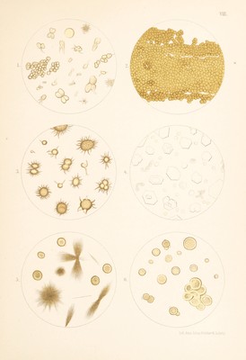
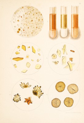
![Fig. 1. SHRED OF TISSUE {part of a tumour) passed with the urine, con¬ sisting of closely packed cells (mostly epithelial) covered with acicular crystals and a few rhombic plates of licematoidin. From a case of CARCINOMA OF THE BLADDER. Fig. 2. Naked eye appearances of some urinary sediments. Middle test tube—White sediment of amorphous phosphates (he., neutral and basic EARTHY PHOSPHATES). Right test tube—PINK URATES. Left test tube—Pale or CLAY-COLOURED URATES. FrG. 3. URIC ACID CRYSTALS. Whet-stone, spindle, barrel, rectangular, and quadratic forms, a few showing narrow rose-coloured bands of urinary pigments, [illustrating irregular modes of growth and pigmentation]. Fig. 4. URIC ACID CRYSTALS in the form of regular and irregular whet¬ stone crystals grouped in various ways. Fig. 5. URIC ACID CRYSTALS in rosettes and sunflower forms. Fig. 6. URIC ACID CRYSTALS. “Drusy forms.” [The word druse employed by the author is more properly applied to a cavity, the walls of which are covered with crystals projecting into the cavity. The forms represented in the picture are composed of a large number of twinned crystals radiating from an axis, and really constitute a small crystalline concretion. This remark applies also to Figs. 4 and 5.]](https://iiif.wellcomecollection.org/image/b29309116_0046.jp2/full/800%2C/0/default.jpg)