Atlas of urinary sediments : with special reference to their clinical significance / by Hermann Rieder ; translated by Frederick Craven Moore ; edited and annotated by A. Sheridan Delépine.
- Hermann Rieder
- Date:
- 1899
Licence: Public Domain Mark
Credit: Atlas of urinary sediments : with special reference to their clinical significance / by Hermann Rieder ; translated by Frederick Craven Moore ; edited and annotated by A. Sheridan Delépine. Source: Wellcome Collection.
50/272
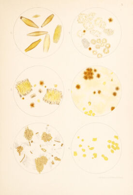
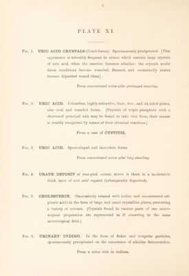
![Fig. 1. URIC ACID CRYSTALS. Flask-like and spindle forms. [Showing the breaking up of large crystals into smaller crystals, an appearance which may be seen in urines undergoing alkaline fermentation. This appearance is also occasionally produced during the process of crystallisation.] Fig. 2. BILIRUBIN. In the form of brownish-red needles arranged in stellate masses scattered about or in the interior of swollen and degenerated cells, which also contain variously coloured granules of biliary pigment. Fig. 3. BILE (ICTERIC) PIGMENTATION OF VESICAL EPI¬ THELIUM. Needles of bilirubin arranged in stellate masses, bile- stained crystals of TRIPLE PHOSPHATE, isolated and grouped in large masses. From a case of CATARRHAL JAUNDICE. Fig. 4. BILE PIGMENTATION OF RENAL EPITHELIUM. Bilirubin in the form of amorphous deposit and acicular crystals, partly free and partly grouped in stellate masses. From a case of CATARRHAL JAUNDICE. Fig. 5. EPITHELIUM FROM THE MALE URETHRA. Cylindrical epithelial cells in groups, and isolated ; the free surface of almost all the cells is stained by blood pigment. From a case of PROSTATITIS. Fig. 6. BILE PIGMENTATION OF EPITHELIUM FROM THE KIDNEY AND URINARY PASSAGES. From the urine of a case of CONGENITAL SYPHILIS with HEPATIC LESIONS and with CHRONIC PARENCHYMATOUS NEPHRITIS. (See also Fig. 4, Plate XXIV.)](https://iiif.wellcomecollection.org/image/b29309116_0050.jp2/full/800%2C/0/default.jpg)