Anatomical atlas : illustrative of the structure of the human body / by Henry H. Smith, under the supervision of William E. Horner.
- Smith, Henry H. (Henry Hollingsworth), 1815-1890
- Date:
- 1845
Licence: Public Domain Mark
Credit: Anatomical atlas : illustrative of the structure of the human body / by Henry H. Smith, under the supervision of William E. Horner. Source: Wellcome Collection.
Provider: This material has been provided by the Woodruff Health Sciences Center Library at Emory University, through the Medical Heritage Library. The original may be consulted at the Woodruff Health Sciences Center Library, Emory University.
16/388
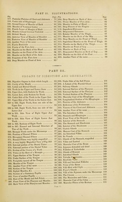
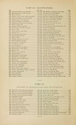
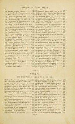
![F,g- Tage ) 187. Posterior Parietes of Chest and Abdomen . 79 '/ 188. Under side of Diaphragm 79 } 189. Second Layer of Muscles of Back 80 j 190. Muscles of Vertebral Gutter 80 191. Fourth Layer of Muscles of Back 81 192. Muscles behind Cervical Vertebrae 81 j 193. Deltoid Muscle 82 194. Anterior View of Muscles of Shoulder . 195. Posterior View of Muscles of Shoulder . 196. Another View of the same 197. Fascia Brachialis 198. Fascia of the Fore-Arm 199. Muscles on the Back of the Hand 83 ] 200. Muscles on the Front of the Arm 84 I 201. Muscles on the Back of the Arm 84 , 202. Pronators of the Fore-Arm 84 203. Deep Muscles on Front of Arm 85 '■ 82 82 82 83 83 Page Deep Muscles on Back of Arm b5 Flexor Muscles of Fore-Arm 86 Muscles in Palm of Hand 86 Deep Flexors of the Fingers 86 Superficial Extensors 87 Deep-seated Extensors 87 Rotator Muscles of the Thigh 87 Muscles on the Bick of the Hip 88 Deep Muscles on the Front of Thigh 88 Superficial Muscles on the Front of Thigh 89 Muscles on the Back of the Thigh 89 Muscles on Front of Leg 89 Muscles on Back of Leg 90 Deep-seated Muscles on Back of Leg 90 Muscles on the Sole of the Foot 90 Another View of the same 90 PART III. ORGANS OF DIGESTION AND GE NE P. A_TI ON. 220. 221. 222. 223. 224. 225. 226. 227. 228 236 244 252 260 266 268. 269. 270. 271. 272. 273. 274. 275. 276. 277. 278. 279. 280. 281. 282. 283. 284. 285 286 287 288 Digestive Organs in their whole length ... 93 ? 289- Cavity of the Mouth 93 290. Labial and Buccal Glands 93 ? 291. Teeth in the Upper and Lower Jaws 94 s 292. Upper Jaw, with Sockets for Teeth 94 293. Lower Jaw, with Sockets for Teeth 94 294. Under Side of the Teeth in the Upper Jaw. 94 295. Upper Side of the Teeth in the Lower Jaw. 94 296. to 235. Eight Teeth, from one side of the 297. Upper Jaw 95 j298. to 243. Eight Teeth, from one side of the 299. Lower Jaw 95 < 300. to 251. Side View of Eight Upper Jaw 301. Teeth 95 302. to 259. Side View of Eight Lower Jaw 303. Teeth 96 304. to 265. Sections of Eight Teeth 96 305. to 267. Enamel and Internal Structure of j 306. Two of the Teeth 96 j 307. Bicuspis Tooth under the Microscope 97 308. Position of Enamel Fibres 97 309. Hexagonal Enamel Fibres 97 310. Enamel Fibres very highly magnified 97 311. A very highly Magnified View of Fig. 268 97 312. Internal portion of the Dental Tubes 98 313. External portion of the Dental Tubes 98 314. Section of the Crown of a Tooth 98 315. Tubes at the Root of a Bicuspis 98 316. Upper Surface of the Tongue 98 317. Under Surface of the Tongue 99 318. Periglottis turned off the Tongue 99 319. Muscles of the Tongue | 32°- Another View of the same 99 321. Section of the Tongue 100 322. Styloid Muscles, &c 10° 323- Section of a Gustatory Papilla 100 j 324. . View of another Papilla 10° j 325- . Root of the Mouth and Soft Palate 101 j 326. . Front View of the Pharynx and Muscles . . 101 ( 327. ' Back View of the Pharynx and Muscles. .. 101 J 328. Page 11. Under Side of the Soft Palate 101 A Lobule of the Parotid Gland 101 Salivary Glands 102 Internal Surface of the Pharynx 102 External Surface of the Pharynx 102 Vertical Section of the Pharynx 103 Muscular Coat of the (Esophagus 103 Longitudinal Section of the OEsophagus . . 103 Parietes of the Abdomen 104 Reflexions of the Peritoneum 104 Viscera of the Chest and Abdomen 105 Another View of the same 105 The Intestines in Situ 106 Stomach and (Esophagus 106 Front View of the Stomach 107 Interior of the Stomach 107 The Stomach and Duodenum 107 Interior of the Duodenum 108 Gastric Glands 1°8 Mucous Coat of the Stomach 108 An Intestinal Villus 108 Its Vessels 108 Glands of the Stomach, magnified 108 Villus and Lacteal ] °8 Muscular Coat of the Ileum 103 Jejunum distended and dried 109 Follicles of Lieberkuhn 109 Glands of Brunner 1 °9 Intestinal Glands 1°9 Valvulse Cc^aniventes 109 Ileo-Colic Valve 109 Villi and Intestinal Follicles HO Veins of the Ileum nQ Villi filled with Chyle HO Peyer's Glands HO Villi of the Jejunum under the Microscope 110 The Caecum Hl The Mesocolon and Colon HI Muscular Coat of the Colon HI Muscular Fibres of the Rectum HI](https://iiif.wellcomecollection.org/image/b21036913_0016.jp2/full/800%2C/0/default.jpg)
 No text description is available for this image
 No text description is available for this image
 No text description is available for this image