Anatomical atlas : illustrative of the structure of the human body / by Henry H. Smith, under the supervision of William E. Horner.
- Smith, Henry H. (Henry Hollingsworth), 1815-1890
- Date:
- 1845
Licence: Public Domain Mark
Credit: Anatomical atlas : illustrative of the structure of the human body / by Henry H. Smith, under the supervision of William E. Horner. Source: Wellcome Collection.
Provider: This material has been provided by the Woodruff Health Sciences Center Library at Emory University, through the Medical Heritage Library. The original may be consulted at the Woodruff Health Sciences Center Library, Emory University.
17/388
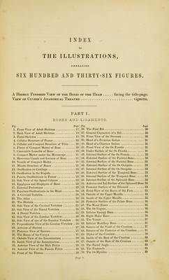
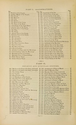
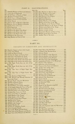
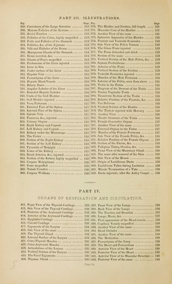
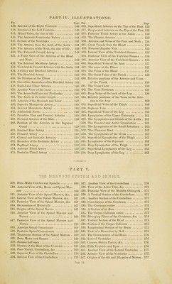
![Fig. 329. 330. 331. 332. 333. 334. 335. 336. 337. 333. 339. 340. 341. 342. 343. 344. 345. 340. 347. 348. 34). 350. 351. 352. 353. 354. 355. 356. 357. 358. 359. 360. 361. 362. 363. 364. 365. 366. 367. 368. 369. Pa Curvatures of the Large Intestine 1 Mucous Follicles of the Rectum 1 Rectal Pouches 1 Follicles of the Colon, highly magnified . . 1 Folds and Follicles of the Stomach 1 Follicles, &c. of the Jejunum 1 Villi and Follicles of the Ileum 1 Muciparous Glands of the Stomach 1 Ileum inverted, *Scc 1 Glands of Peyer magnified 1 Peritoneum of the Liver injected 1 Liver in Situ 1 Under surface of the Liver 1 Hepatic Vein 1 Parenchyma of the Liver 1 Hepatic Blood-Vessels 1 Biliary Ducts 1 Angular Lobules of the Liver 1 Rounded Hepatic Lobules 1 Coats of the Gall Bladder 1 Gall Bladder injected 1 Vena Portarum 1 External Face of the Spleen 1 Internal Face of the Spleen 1 Splenic Vein 1 Pancreas, &c, injected 1 Urinary Organs 1 Right Kidney and Capsule 1 Left Kidney and Capsule 1 Kidney under the Microscope 118 The Ureter 118 Section of the Right Kidney 118 Section of the Left Kidney 1 Pyramids of Malpighi 119 Lobes of the Kidney 119 Renal Arteries, &c., injected 119 Section of the Kidney highly magnified . . 120 Corpora Malpighiana 120 Same magnified \OQ Tubuli Urinifcri 120 Corpora Wolfiana 120 Fig. 370. 371. 372. 373. 374. 375. 376. 377. 378. 379. 380. 5 381. s382. i 383. |384. ^385. \ 386. 1387. 5388. 1389. 5390. J391. j 392. 393. | 394. ^395. | 396. 1397. | 398. i 399. 1400. Uoi. 402. 403. 404. 405. 406. 407. 408. 409. 410. Page The Bladder and Urethra, full length 121 Muscular Coat of the Bladder 121 Another View of the same 121 Sphincter Apparatus of the Bladder 122 Prostate and Vesiculae Seminales 122 Side View of the Pelvic Viscera 122 The Glans Penis injected 123 The Penis distended and dried 123 Section of the same 123 Vertical Section of the Male Pelvis, &c. .. 123 Septum Pectiniforme 123 Arteries of the Penis 123 Vertical Section of the Urethra 123 Vesiculae Seminales injected 123 Muscles of the Male Perineum 124 Interior of the Pelvis, seen from above ... 124 Testis in the Foetus 125 Diagram of the Descent of the Testis .... 125 Tunica Vaginalis Testis 125 Transverse Section of the Testis 125 Relative Position of the Prostate, &.c 126 Vas Deferens ,. . 126 Vertical Section of the Bladder 126 The Testicle injected with Mercury 126 Another View 126 Minute Structure of the Testis 126 Female Generative Organs 127 Another View of the same 127 External Organs in the Foetus 127 Muscles of the Female Perineum 127 Side View of the Female Pelvis, &c 128 Relative Position of the Female Organs .. 128 Section of the Uterus, &,c 128 Fallopian Tubes, Ovaries, &c 129 Front View of the Mammary Gland 129 The same after removal of the Skin 129 Side View of the Breast 130 Origin of Lactiferous Ducts 130 Lactiferous Tubes during Lactation 130 Minute Termination of a Tube 130 Ducts injected; after Sir Astley Cooper . . 130 PART IV. ORGANS OF RESPIRATION AND CIRCULATION. 411. Front View of the Thyroid Cartilage 133 j 427. 412. Side View of the Thyroid Cartilage 133 J 428. 413. Posterior of the Arytenoid Cartilage ] 33 I 429. 414. Anterior of the Arytenoid Cartilage .. 133 430. 415. Epiglottis Cartilage 133 I 431. 416. Cricoid Cartilage 133 > 432. 417. Ligaments of the Larynx 134 ' 433. 418. Side View of the same 134 434. 419. The Thyroid Gland ] 34 435. 420. Internal Surface of the Larynx 134 436. 421. Crico-Thyroid Muscles 135 437. 422. Crico-Arytenoid Muscles 135 438. 423. Articulations of the Larynx 135 439. 421. Vertical Section of the Larynx 135 \ 440. 425. The Vocal Ligaments 1 35 ' 411. 426. Thymus Gland 135 | 442. Front View of the Lungs 136 Back View of the Lungs . 136 The Trachea and Bronchia 137 Lungs, Heart, &c 137 First appearance of the Blood-vessels 138 Capillary Vessels magnified 138 Another View of the same 138 Blood Globules 138 Another View of the same 138 The Mcdiastina 139 Parenchyma of the Lung 139 The Heart and Pericardium 139 Anterior View of the Heart 139 Posterior View of the Heart ] 39 Anterior View of its Muscular Structure .. 140 Posterior View of the same ... 140](https://iiif.wellcomecollection.org/image/b21036913_0017.jp2/full/800%2C/0/default.jpg)
 No text description is available for this image
 No text description is available for this image
 No text description is available for this image